Our dogs use their paws for digging, holding things, playing, demanding our attention and scratching their little doggy ears and faces. Given the amount of dirt and germs they can track on their feet after each walk, it's no wonder that they can often get eye infections.

Some of the common dog eye problems affect the eyelids, eyelashes and the lens of the eye. Symptoms can range from excessive tearing to sudden blindness - as in the case of inflammation of the optic (eye) nerve.

While some eye conditions in dogs can be treated easily with artificial tears and flushing the eye out with lukewarm water, there are others that require medical and even surgical interventions.

These eye problems can happen to any dog. However, there are some breeds that are more prone to some conditions. For example, pugs are more prone to conditions like entropion (in which a section of the eyelid is folded inward) and conjunctivitis. Other risk factors include genetic factors and previous injury or medical condition.

Read on to know about the most common dog eyelid problems, dog eyelash problems, glaucoma and cataract in dogs, cherry eye in dogs, keratoconjunctivitis in dogs and conjunctivitis in dogs. The symptoms, causes, risk factors, diagnosis and treatment, including surgery, for these conditions have also been discussed below.

  1. Dog eyelid problem: entropion
  2. Dog eyelid drooping: ectropion
  3. Lagophthalmos: dog can't close eyes fully
  4. Conjunctivitis in dogs
  5. Dog eyelid problems: blepharitis
  6. Dog eyelash problems: distichiasis, trichiasis and ectopic cilia
  7. Glaucoma: blindness in dogs
  8. Cloudy eyes in dogs: old age and cataracts
  9. Progressive retinal atrophy (PRA): blindness in dogs
  10. Cherry eye in dogs
  11. Keratoconjunctivitis sicca (KCS) or dry eye in dogs
  12. Dog can't see suddenly: optic neuritis

Entropion is a condition in which a part of the dog's eyelids is curled or folded inwards. This can lead to symptoms such as

  • Discomfort or pain as the eyelashes and fur can irritate the cornea.
  • This, in turn, can cause your dog's eyes to become red and teary.
  • The dog may also squint a lot or keep his or her eyes shut most of the time
  • Or, have abnormal discharge from the eyes.

In some cases, there are no complications or pain and the condition doesn’t attract medical attention. However, some dogs will face discomfort and pain and will constantly paw at and rub their eyes. They will also constantly wink in an attempt to relieve the irritation in their eyes. In some cases, there is a mucus-like discharge from the eyes. The hair around the eyelids may appear damp and matted more than usual.

Causes and risk factors: The biggest risk factor for entropion is genes. In cases where entropion is inherited from the dog parents, it is often seen in younger dogs below the age of one. Less often, secondary factors cause entropion. These may be trauma to the eye, eye infection, corneal spasms or neurological issues.

Certain breeds are predisposed to it owing to the structure of their faces: this condition is more common in Boxers, Cocker Spaniels, English Bulldogs, Golden Retrievers, Great Danes, Irish Poodles, Pugs and St Bernards.

Diagnosis: Diagnosis is usually straightforward: if no obvious facial abnormalities or serious underlying causes are noted, the vet will conduct a physical exam of the eye.

Treatment: Topical antibiotics may be prescribed to combat any infections. In severe case, corrective surgery - known as blepharoplasty - may be needed to remove the excess skin from the eyelid and tighten the eye socket.

Prognosis: The prognosis is usually good and the likelihood of a relapse is quite low.

Complications: If the condition worsens, ulcers can form on the cornea (eye lens) and there may be consequences for the dog's eyesight. Eye infection, scarring of the eye and corneal perforation are also possible. Pigment deposits on the eye can also lead to vision problems.

Ectropion is a common eye problem in dogs. It occurs when the lower eyelid droops outwards and makes the lower part of the eye appear red and bloody. Normally, the eyelid acts as a physical barrier to dirt and air, so this deformity makes the dog more likely to contract conjunctivitis and a host of other infections.

The symptoms include:

  • Droopy eyes
  • Redness in the lower part of the eye
  • Tearing that causes a pigment stain near the eyes and nose of the dog
  • A mucoid discharge from the eyes
  • Irritation in the eyes, which may cause the dog to paw at or rub the eyes frequently.
  • Ectropion can be painful for the dog.
  • Scarring on the cornea, known as keratitis, may occur in more severe cases.

Causes: The condition is thought to have a genetic component to it which makes the muscles around the eyes a little weaker. Certain breeds such as Cocker Spaniels, Saint Bernards, Mastiffs and Bloodhound are predisposed to the condition.

Sometimes there may be an underlying cause (secondary cause). Acquired ectropion, as it is called, may be caused by facial nerve paralysis, hypothyroidism, or neuromuscular disease.

Complications: Dogs with this condition are likely to develop conjunctivitis and keratitis. 

Diagnosis: It is usually diagnosed in younger dogs before the age of one. For dogs who develop this condition later in life, a secondary cause is more likely to behind it. If other symptoms are absent, the diagnosis can be made quite simply by conducting a physical examination. If corneal damage or ulceration is suspected, corneal staining will be conducted. If systemic issues are at play, then more detailed blood work and imaging tests will be carried out as well.

Treatment: Some mild, ophthalmic antibiotics may be prescribed to ward off infections. Artificial tears or similar lubricating agents may be suggested to keep the eye moist. A cold or warm compress may ease some inflammation and discomfort.

The surgical route is taken in more severe cases that do not respond to medication. The eyelid is sutured closer to the eye to correct the defect.

Prognosis: The prognosis is usually good as relapse does not occur often. However, there is a chance of developing entropion if there is an overcorrection. If there is no corneal impairment, then surgery is usually curative.

Complications: If the problem persists, more serious issues can develop. Since the eyelids provide lubrication and protection to the eyes, impairment in their functioning will make the eyes more susceptible to infection. Corneal inflammation may set in followed by ulceration. In severe cases, the dog could run the risk of going blind.

Lagophthalmos is the inability to fully close the eyelids. This is largely due to genetic reasons and brachycephalic dogs (those with short noses and flat faces like pugs) are disproportionately affected.

Symptoms: The classic symptom of lagophthalmos is the inability to fully close the eyes even during sleep.

Diagnosis: If you notice a space between your dog’s eyelids when he or she is asleep, it could indicate that your dog suffers from this condition. Take your dog to a vet for a confirmed diagnosis. If there are no accompanying symptoms, diagnosis can be made by observation. If underlying causes are suspected, imaging tests and blood samples will be taken. The intraocular (within the eye) pressure will also be checked to investigate localized lesions. 

Treatment: Warm or cold compresses may ease some of the irritation, and artificial tears may be recommended by the vet to keep the eyes lubricated. Your doctor may advise medicines to avoid infection. Surgery is usually recommended for dogs who are in a stable condition and such dogs are likely to benefit from it. The procedure is called the lateral or medial canthoplasty and involves removing a part of the eyelid to give it more of an almond shape. This makes it easier for the dog to close fully the eyes.

Prognosis: The surgery generally has a good prognosis and the dog should not have a recurrence.

Conjunctivitis is the inflammation of the conjunctiva - a thin, clear membrane within the eye that stretches across the surface and lines the inside of the eyelid. Its role is protective as it traps dust and debris and prevents them from causing infections. It also facilitates smooth movement of the eyelid.

Symptoms: Conjunctivitis in dogs looks similar to conjunctivitis (pink eye) in humans. The symptoms include:

  • Reddening of the eye
  • Excessive tear production
  • Swelling of the area around the eye
  • Pain

Dogs with this eye condition may paw at their eyes constantly to relieve the pain. 

Causes: Many different issues, including environmental allergens, congenital factors, viral infections, trauma or secondary underlying issues, can cause conjunctivitis in dogs. 

Diseases such as dry eye, ulcerative keratitis, anterior uveitis, glaucoma, diseases of the eyelid and eyelash, and certain cancers can all precipitate conjunctivitis.

Risk factors: Facial structure can predispose certain breeds to the condition. Flat nosed dogs (Pugs, Bulldogs) are especially prone.  

Diagnosis: Since the condition can be caused by several underlying issues, your vet will ask for a detailed medical history of your dog and check for other symptoms. Since blockages in the nasal cavity can also cause this condition, your dog's nose may be examined as well. The pressure and tear production of the eye could come under scrutiny, too.

Treatment: Treatment will depend on the diagnosis. Since the cause is often pathogenic, appropriate antibiotics and medicines will be prescribed. Cultures will be taken if parasitic or viral causes are suspected. For cancerous growths, treatment will become more involved and will require further treatment. In an otherwise healthy dog, however, the condition should pass within a few days - check with your doctor about eye drops to clean the eye and relieve the pain and swelling.

Prognosis: The prognosis is generally very good.

Complications: Issues with the conjunctiva need to be addressed since neglect can lead to blindness. As with anything symptomatic, the underlying cause can be more serious than a generalized flare up.

Inflammation of the area around the eyelashes and within the eyelashes (connective tissues and glands) is known as blepharitis. This condition can affect one or both the eyelids.

Symptoms: A painful condition, blepharitis may present as

  • Swelling, redness and itchiness of the eyelid
  • Some dogs may also experience blepharospasm: spasmodic blinking
  • Pawing or scratching the eyes may be a sign of irritation and pain
  • Discharge from the affected eye that can turn into a crusty deposit around the eyelid

Causes: The causes of blepharitis can range from an underlying health condition to proximate causes like allergic reactions caused by mites, ticks, certain foods and drugs, bacterial infections caused by pathogens such as staphylococcus and streptococcus, certain cancers such as sebaceous adenomas, trauma to area around the eye, parasitic infection such as mange and idiopathic (or unknown) reasons.

Risk factors: Dogs with bulging eyes, narrow muzzles and skin folds on the face are more prone to this condition.

Diagnosis and treatment: Diagnosis will be based on the clinical history you provide, so make sure to be thorough. Since systemic issues may be responsible and blepharitis could be merely coincidental, your vet will likely conduct imaging and blood tests. Treatment will be designed to address symptoms and will be contingent on diagnosis.

Distichiasis

Distichiasis is characterised by excessive eyelash hair that grows from the meibomian gland, from which hair should not normally grow.

Symptoms: The extra eyelashes are often stiff and constantly prick the dog's eyes. Symptoms include:

  • Excessive tearing
  • Squinting
  • Blinking
  • Your dog may try to keep his or her eyes closed if the pain becomes severe

Causes: Distichiasis is a genetic disease; that is, it is inherited from your pet's dog parents. According to veterinary reports, it is one of the most commonly reported eye issues among dogs.

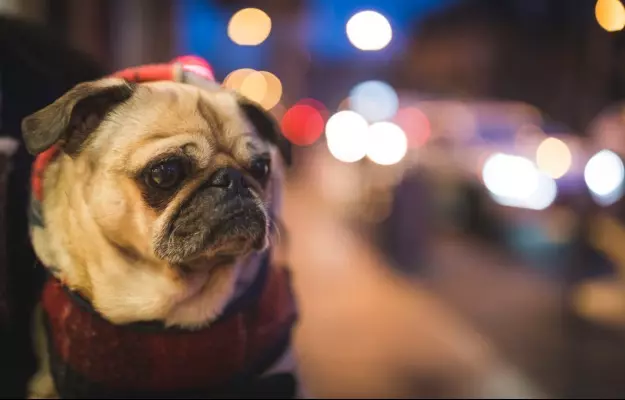
Risk factors: While distichiasis can happen to any dog at any age, it is most commonly seen in younger dogs below the age of three. It is more commonly observed in certain breeds: Cocker Spaniel, Poodle, Pug, and Dachshund are some examples.

Diagnosis: Take your doctor to the vet for a physical examination of the eye if he or she exhibits the symptoms of distichiasis. If your dog's extra eyelash hairs are very fine, diagnosis may be more challenging. But usually, the excess hair can be examined easily and indicated as the cause of the issue.

Treatment: Surgical means are necessary to get rid of distichiasis. Plucking the eyelashes is not permanent as they will grow back. The goal of the surgery is to eradicate the hair follicles causing the hair to grow. While it is not a very involved procedure, the dog may need to be sedated with general anaesthesia as the area is sensitive and any sudden movements of the head can cause damage.

Prognosis: While the prognosis from the surgery is usually good, the eyelashes may grow back if some of the follicles are missed. This means that a follow-up procedure may become necessary.

Complications: Similar to other conditions, underlying causes could exacerbate distichiasis. If that is the case, there will be other symptoms. You must take your dog to the vet if you suspect that there is constant inflammation in the eyes. This is because home remedies to combat distichiasis are short term and not very effective. If irritation persists, the cornea can get ulcerated (small cracks in the eyeball) and the complications will then become harder to resolve.

Trichiasis

In this condition, the dog has ingrown eyelashes or eyelashes growing the wrong direction (towards the eye). However, unlike distichiasis, the eyelash growth in trichiasis is not from an abnormal spot.

Symptoms: The problems related to dogs' eyelashes have similar symptoms:

  • Epiphora (excessive tears)
  • Change in iris pigmentation - colour of the eyeball
  • Blepharospasm (twitching of the eye)
  • Eye infection

Causes and risk factors: Dogs with long facial hair like Shih-Tzus, multiple folds of skin like Bulldogs and brachycephalic dogs like pugs are more prone to developing this condition.

Diagnosis: The Schirmer tear test, in which filter paper is placed on the lower eyelid and kept in place for 5 minutes to assess the level of tear production, may be administered. The eyes may be stained with fluorescein to test for ulcers. 

Treatment: Surgical intervention, similar to that involving distichiasis, is usually required to solve this issue. The doctor may recommend cryosurgery to target the hair follicles or remove a part of the eyelid to prevent further hair growth in the wrong direction.

Complication: The ingrown hairs can irritate the eye and even scratch the cornea. If left untreated, the condition may lead to corneal ulcers and keratitis (inflammation of the cornea.

Ectopic cilia

In this condition, the hair grows from inside the conjunctiva - inner part of the eyelid. 

Symptoms: Like the other two eyelash conditions in dogs, ectopic cilia also causes irritation and pain in the eyes, excessive tear production and eyelid twitching.

Causes and risk factors: Dogs with long hair, brachycephalic faces and excessive skin folds present with this condition more often than other dogs. At risk are breeds such as Shih-Tzus, Lhasa Apsos, Pekingese, Retrievers, Collies, Bulldogs and the Boston Terrier.

Diagnosis and treatment: Diagnosis is usually done by physical examination of the dog's eye. Surgery is the usual treatment option - cryosurgery cools the area to remove a little bit of tissue along with the hair to avoid regrowth.

Glaucoma is caused by increased pressure within the eye. The increased intraocular pressure (IOP) can cause damage to the retina and optic nerve and eventually lead to blindness.

The increased pressure builds up because of inadequate drainage of fluid. Fluid drains from the space between the cornea and iris: this area is known as the iridocorneal or drainage angle.

Symptoms: The symptoms of glaucoma are subtle initially, which makes it hard to diagnose in the early stages. Certain breeds are more susceptible to glaucoma and purebred dogs are also known to be disproportionately affected. The symptoms include: 

  • Vision loss
  • Cloudiness at the front of the eye
  • Enlargement of the eyeball
  • Inflammation of the eye
  • Headaches
  • Lethargy in dogs
  • Change in behaviour
  • Acute glaucoma can be very painful due to the sudden increase in pressure and there will be an impairment in vision as well. 

Types and causes: In primary glaucoma, pressure builds on the eye due to the improper development of the drainage angle. Secondary glaucoma, on the other hand, may be caused by lens luxation (which is the movement of the lens within the eye), localized tumours, trauma, blood clots that prevent the drainage of fluid or a generalized inflammation of the eye tissue. The danger of glaucoma is that even with treatment, 40% of dogs will go blind. 

Diagnosis: As with other eye conditions, your vet will ask for a detailed history as underlying issues may be causing the condition. A tonometer, which gauges pressure within the eye, will be used to check if there is increased pressure since this development can lead to irreversible damage.

Treatment: The immediate aim of treatment is to relieve this excess pressure. Depending on the severity of the condition, your vet will prescribe a number of drugs to lower the pressure in the eye.

Alternatively or additionally, your vet may suggest cyclocryotherapy. This procedure uses extremely cold temperatures to kill fluid-producing cells in the eye, to arrest the progression of the disease.

However, fluid-reducing drugs and techniques are not very effective in dogs whose vision has already been affected due to the degeneration of the optic nerve. In such cases, the vet may suggest removing the eye to relieve any more pain in the area.

Age-related lenticular sclerosis

Normally, the lens of the eye focuses incoming light on to the retina. The retina then sends signals to the brain, which perceives what we see. The lens can dilate or constrict to control the amount of light entering in a process called accommodation. As dogs age, the eye lens and muscles deteriorate. This can lead to lenticular sclerosis: if you have noticed a bluish tinge in the eyes of older dogs, then you have seen the biggest sign of this condition. Lenticular sclerosis is not painful for dogs, although it can affect their vision slightly. Usually, this condition requires no treatment.

Cataract

Cataract, on the other hand, occurs because of abnormal clumping of proteins on the lens of your pets' eyes.

Symptoms: Cataract symptoms include cloudy vision. A portion of the lens can become opaque, which means it can no longer perceive light.

Cataracts are distinguished by severity. If there is less than 30% of opacity of the lens, then there will hardly be any symptoms and the dog will be able to see and function normally. For cataracts with over 60% coverage, the dog may show symptoms such as vision loss and inability to see things in dim settings.

Causes and risk factors: Sometimes cataracts are congenital, or they can be influenced by genes. They may also be caused by trauma, or occur over time with age. Electric shocks and dietary deficiencies can also lead to cataracts in dogs. The most commonly affected breeds are Cocker Spaniels, Poodles, Golden Retrievers and Huskies, to name a few.

Diabetic dogs are also highly likely to have cataracts. Increased glucose in the blood means a higher production of sorbitol in the eye. Sorbitol attracts water and causes irreversible cataracts.

Uveitis, which is an inflammation of the middle layer of the eye - this can also lead to cataracts. 

Diagnosis and treatment: In determining the course of the treatment, your vet may order an electroretinogram and ultrasound to ascertain the extent of the damage. An electroretinogram can be used to check the functionality of the retina; if the retina is damaged beyond repair, then there may be no point in surgery unless it is performed to mitigate pain or accompanying issues such as glaucoma.

Cataracts are only approached invasively if there is a good chance of saving the eye. That said, some dogs can live quite normally with blindness in one eye as it may happen gradually and their sense of smell and hearing accommodates for the loss.

Previously, surgical removal was the treatment of choice for dogs who were considered fit enough to withstand anaesthesia and surgery.

The new treatment is called phacoemulsification. It uses sound waves to breach and slowly break down or remove the affected lens. An artificial lens is put in as a replacement. This procedure has been shown to be quite effective.

Prognosis: If cataracts are operated on in the earlier stages, the prognosis is better since they are softer and easier to access. Pre-operation inflammation is also a consideration since the outcome is better without it. There are complications with surgery, including the possibility of deep eye infections and glaucoma. As mentioned above, the decision to opt for surgery is contingent on the overall health of the dog.

Read more: Diabetes in dogs

PRA describes a cluster of congenital conditions that affect the development of the retina. The photoreceptor cells in the retina stop functioning prematurely, and eventually, the dog will lose the ability to see.

Symptoms: The disorder usually begins with night blindness and progresses to inability climbing stairs and walking into objects.

While it may sound like a curse, PRA does not cause any pain or distress to the pet unless there are accompanying conditions as well. Vision loss is usually gradual, which allows the dog time to sharpen his or her other sensory facilities. Given that their sense of smell and hearing is stronger than their ability to see, some dogs will adapt well to the changes. 

In some cases, onset is faster and sight will be compromised at around two months of age and young dogs will be completely blind by two years. In other cases, which are more common, the eyesight will deteriorate gradually and older dogs will lose vision completely. This depends on the nature of the disease; in photoreceptor dysplasias, photoreceptors are faulty at inception and don’t develop properly. The sight will be lost faster in this case. In photoreceptor degenerative disorders, the loss is gradual as the photoreceptor cells dry after formation.

Causes and risk factors: PRA is an inherited disease for which there is no treatment or cure. It is recommended not to breed dogs that are diagnosed with PRA given its genetic development.

Purebreds have been known to have a greater incidence of these conditions. Certain breeds such as Cocker Spaniels, labrador retrievers, and poodles are also predisposed.

Management: With this condition, the challenge is management. While it is upsetting to have a dog lose his or her sight, the owner is likely to be more distressed than the dog.  

If your dog does lose his or her eyesight, make sure that your home is accident-proof. Don’t move furniture around too much, keep food bowls in familiar locations, and install doors on stairs to save your dog from falling down the steps.

Your pet dog has a third eyelid gland that keeps the eye lubricated, along with the tear gland. This gland is normally hidden from view, in the lower part of the eye. When the muscle fibres holding this gland in place become weak, it can cause this gland to prolapse or popout - you see it as a red cherry-like protrusion near the bottom eyelid.

Symptoms: The red protrusion from the eye will be unmissable in dogs with this condition. 

Causes and risk factors: The exact cause of cherry eye in dogs is not known, but it is thought that genes may be responsible for weak eye muscle fibres in some dogs. Cherry eye is mostly seen in dogs below two years of age, and smaller dogs such as Cocker Spaniels, Beagles, Lhasa Apsos, Shih Tzus.

Diagnosis and treatment: Your vet will conduct a thorough physical examination to rule out conjunctivitis. The treatment for cherry eye usually involves surgery.

Prognosis: The prognosis is medium at best. Dogs who get a third eye gland prolapse once are likely to have it again. A surgical procedure called tucking uses a permanent stitch to hold the gland in place. In dogs who have had this surgery two or three times and are at high risk of relapse, the surgeon may prescribe imbrication (cutting a part of the gland) or a combination of imbrication and tucking surgery.

Keratoconjunctivitis sicca in dogs (KCS) is inflammation of the tear gland, conjunctiva and cornea of the eye. The condition is caused by inadequate tear production or a reduction in the water content of the tear film that covers the eye.

Symptoms: Dry eyes can cause redness and irritation in the eyes. Some of the major symptoms of KCS include:

  • Since tears lubricate the eyes and act as an antiseptic, lower production of tears will lead to pain and inflammation. 
  • Dogs with KCS may have a mucoid discharge from the eye, constantly scratch their eyes and blink.
  • The eyes may appear swollen (chemosis) and the colour of the eyes may become yellowish as well.
  • In some cases, there will be cherry eye (prolapse of the third eyelid gland) with a bloody lump present in the lower corner of the eye.
  • In severe cases, blindness may eventually occur.

Causes and risk factors: KCS is quite a common occurrence in dogs, and some causes may be inherited. Certain breeds such as Cocker Spaniels, Shih Tzus and Bulldogs are more prone to it.

Most commonly, KCS is caused by immune-mediated destruction of the tear-producing gland tissue. The progression of this condition is poorly understood, but the body attacks the tear-producing glands and impedes their functioning. 

For dogs with cherry eye, treatment can sometimes involve removing the third eyelid gland. Since this gland makes 30% of the water content in tears, its removal can lead to KCS.

Canine distemper is also known to cause KCS. 

In some breeds that are predisposed, there may be a congenital disorder which affects the development of tear-producing glands, which means there is not as much production.

Certain drugs, including NSAIDs (painkillers), may temporarily lower tear production. Anaesthesia can also affect production for a brief period of time.

Additionally, trauma to the area can damage regular functioning as well.

Treatment: Since immune-mediated destruction is the most common cause, drugs that address this issue are the most commonly employed treatment. Cyclosporine, an immunomodulating drug, lowers this function and has proven to be an effective treatment in most cases.

Artificial tears and moistening agents are common therapies as they ease inflammation and relieve some symptoms. Massaging the area and putting a warm compress on it is also a common home remedy.

Surgery is reserved for severe cases.

The optic nerve transmits visual information from the retina to the brain. Optic neuritis is inflammation and swelling of the optic nerve.

Symptoms: Common symptoms include dilated pupils and sudden blindness. The dog may also experience pain.

Causes: Usually, there is an underlying cause for this condition. Primary optic neuritis is quite rare and seen in younger dogs below the age of three. Central nervous system (CNS) function and certain cancers are common underlying causes.

Diagnosis: A professional opinion is needed to diagnose optic neuritis as it shares the symptoms of other eye disorders. 

Since an underlying cause involves the CNS, the vet may request an analysis of the cerebrospinal fluid. MRI testing may be requested as well. If a bacterial or fungal infection is suspected, there may be a need for blood and urine tests.

Treatment: Since underlying causes make up the majority of these cases, treatment will depend on them. Check with your doctor for pain management options.

References

  1. MSD Veterinary Manual [Internet]. Merck & Co., Inc.; Eye Disorders of Dogs
  2. Tufts Now. [Internet]. Tufts University. Massachusetts; Spotting Eye Problems in Pets
  3. Cathryn Mellersh. The genetics of eye disorders in the dog. Canine Genet Epidemiol. 2014; 1: 3. PMID: 26401320.
  4. Veterinary Partner. [Internet]. Veterinary Information Network. Davis, California; Entropion in Dogs
  5. Canine Lens: Cataract, Luxation, and Surgery. In Essentials of Veterinary Ophthalmology, K.N. Gelatt (Ed.), August 2014.
  6. Vestre W.A. Use of cyclocryotherapy in management of glaucoma in dogs.. Modern Veterinary Practice, February 1984; 65(2): 93-7.
Read on app
cross
Ask your health query from live doctors now!