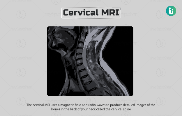
What is a cervical MRI scan? 

The cervical MRI (magnetic resonance imaging) uses a magnetic field and radio waves to produce detailed images of the bones in the back of your neck called the cervical spine. 

During the test, the MRI machine creates a temporary magnetic field in the individual’s body, forcing hydrogen atoms present in the body to align with the field. The transmitter of the machine then sends radio waves that knock the atoms out of alignment. When the radio waves are shut off, the atoms realign and send out distinct signals which are picked up by the receiver of the machine to make detailed images of the scanned area.

  1. Why is a cervical MRI done?
  2. Who cannot have a cervical MRI?
  3. How should I prepare for a cervical MRI?
  4. How is a cervical MRI done?
  5. How will a cervical MRI feel like?
  6. What do the results of a cervical MRI mean?
  7. What are the risks and benefits of a cervical MRI?
  8. What happens after a cervical MRI?
  9. Contrast vs non-contrast cervical MRI
  10. What are the other tests that can be done with a cervical MRI?

Your healthcare practitioner may order this test if you show the following symptoms:

  • Severe pain in the neck, arm or shoulder that does not subside after treatment
  • Neck pain with symptoms like leg weakness or numbness

The test may also be done to detect conditions such as:

The test may also be done before or after spine surgery.

myUpchar doctors after many years of research have created myUpchar Ayurveda Urjas Capsule by using 100% original and pure herbs of Ayurveda. This ayurvedic medicine has been recommended by our doctors to lakhs of people for sex problems with good results.
Long Time Capsule
₹719  ₹799  10% OFF
BUY NOW

Most people can safely undergo an MRI. However, this test may not be recommended if you have any of the following:

  • Pregnancy: MRI scans are not usually recommended during pregnancy.
  • Metal implants or fragments: You may not be allowed to undergo the test if you have metal implants or fragments. However, the medical staff will decide the safety of a scan on a case-by-case basis, as it is possible to make some implants (such as pacemakers) MRI-safe.
  • Tattoos: Some tattoos contain traces of metal due to which you may feel discomfort or heat in your tattoo during the scan.

You may be instructed not to eat/drink for up to six hours before the test. Please inform your healthcare practitioner and the radiologist if you have any of the following:

  • Heart pacemaker/defibrillator
  • Ear implants
  • Programmable shunts
  • Stents (not in the heart)
  • Electronic/implanted stimulators or devices
  • Implanted drug pumps
  • Aneurysm clips and coils
  • Filters, such as blood clot filters
  • Metal fragments in the body, like bullets, shrapnel or other metal pieces
  • Pregnancy
  • Unable to lie on the stomach for an hour

If you have a fear of closed spaces, you may be given a mild sedative that will make you sleepy and less anxious.

Leave all jewellery and valuables like keys, watches, etc. at home, as they cannot be taken inside the MRI room.

An MRI scan usually involves the following steps:

  • The radiographer will instruct you to remove all metal objects, including dentures and wigs, before entering the scan room.
  • They may ask you to wear a hospital gown.
  • You will have to lie on a bed, which will slide into the MRI scanner.
  • The procedure may require a dye (contrast), which will be given through a vein in your arm before the test. The contrast helps to see certain areas more clearly.
  • You will have to lie very still, as movements can blur/distort the pictures. The test usually lasts for 30-60 minutes but can take longer.
myUpchar doctors after many years of research have created myUpchar Ayurveda Kesh Art Hair Oil by using 100% original and pure herbs of Ayurveda. This Ayurvedic medicine has been recommended by our doctors to more than 1 lakh people for multiple hair problems (hair fall, gray hair, and dandruff) with good results.
Bhringraj Hair Oil
₹599  ₹850  29% OFF
BUY NOW

The procedure generally causes no pain. You may feel that the bed is hard or cold. If so, you can ask for a blanket or pillow. The machine makes loud noises during the procedure, but you will be provided earplugs to block out the noise. You may feel some warmth during the test, which is completely normal.

If a contrast dye is given to you during the test, you may feel a cool sensation as it is being injected into the intravenous line.

A normal result indicates that the part of the spine and nearby nerves present in the neck appear normal. 

Abnormal results may be due to:

  • Cervical radiculopathy (slipped disc)
  • Cervical spondylosis (abnormal wear of the bones and cartilages in the neck)
  • Spinal stenosis (narrowing of the cervical spine)
  • Osteomyelitis (infection of the bone)
  • Infection of the spine
  • Spinal cord injury or compression
  • Spinal tumour
  • Degenerative changes due to age
  • Discitis (inflammation of the disc)
  • Multiple sclerosis (inflammation of the covering of nerves)
  • Spinal fracture

The following risks are associated with this test:

  • Allergic reaction to the contrast (which is rare).
  • Malfunction of metal implants: The strong magnets present in the MRI machine may cause pacemakers and other metal implants to malfunction or move/shift inside the body. 
  • The contrast used may be harmful to people with kidney diseases that require dialysis.

The benefits associated with the test include:

  • Generally painless
  • Does not use radiation
  • Gives detailed diagnostic pictures
myUpchar doctors after many years of research have created myUpchar Ayurveda Urjas Energy & Power Capsule by using 100% original and pure herbs of Ayurveda. This Ayurvedic medicine has been recommended by our doctors to lakhs of people for problems like physical and sexual weakness and fatigue, with good results.
Power Capsule For Men
₹719  ₹799  10% OFF
BUY NOW

You will be able to leave soon after the test and continue with your daily activities. However, if a sedative was given:

  • You must not drive a car or use public transport.
  • Make sure you have someone with you for 24 hours after the test is performed.
  • Avoid operating machinery on the day of the test.

A contrast dye may be administered during the MRI to improve the clarity of certain areas.c The dye is usually used in conditions such as suspected severe soft tissue injury as these cannot be seen on an MRI without contrast. Contrast is not necessary for the diagnosis of conditions like oedema, hematoma, etc.

The dye will be administered as an injection or through a vein in your arm. When the technician injects the contrast, you may feel a cool sensation. In rare cases, a person may have an allergic reaction to the dye. If a person has kidney disease and requires dialysis, the contrast may be harmful to them.

Additional tests that may be conducted along with this test include:

Disclaimer: All results must be clinically correlated with the patient’s complaints to make a complete and accurate diagnosis. The above information is provided from a purely educational point of view and is in no way a substitute for medical advice by a qualified doctor.

References

  1. Nemours Children’s Health System [Internet]. Jacksonville (FL): The Nemours Foundation; c2017; Magnetic Resonance Imaging (MRI): Cervical Spine
  2. US Food and Drug Administration [Internet]. Maryland. US; MRI (Magnetic Resonance Imaging)
  3. National Health Service [Internet]. UK; MRI scan
  4. Chou R, Qaseem A, Owens DK, Shekelle P; Clinical Guidelines Committee of the American College of Physicians. Diagnostic imaging for low back pain: advice for high-value health care from the American College of Physicians. Ann Intern Med. 2011;154(3):181-189. PMID: 21282698
  5. Gardocki RJ, Park AL. Degenerative disorders of the thoracic and lumbar spine. In: Azar FM, Beaty JH, Canale ST, eds. Campbell's Operative Orthopaedics. 13th ed. Philadelphia, PA: Elsevier; 2017:chap 39
  6. Wilkinson ID, Graves MJ. Magnetic resonance imaging. In: Adam A, Dixon AK, Gillard JH, Schaefer-Prokop CM, eds. Grainger & Allison's Diagnostic Radiology: A Textbook of Medical Imaging. 6th ed. Philadelphia, PA: Elsevier Churchill Livingstone; 2015:chap 5
  7. Even JL, Eskander MS, Donaldson WF. Cervical spine injuries. In: Miller MD, Thompson SR, eds. DeLee and Drez's Orthopaedic Sports Medicine. 4th ed. Philadelphia, PA: Elsevier Saunders; 2015:chap 126
  8. Koerner JD, Vaccaro AR. Evaluation, classification, and treatment of cervical (C3-C7) injuries. In: Winn HR, ed. Youmans and Winn Neurological Surgery. 7th ed. Philadelphia, PA: Elsevier; 2017:chap 306
  9. Cleveland Clinic [Internet]. Ohio. US; Magnetic Resonance Imaging (MRI)
  10. Better health channel. Department of Health and Human Services [internet]. State government of Victoria. Australia; MRI scan
  11. Michigan Medicine [internet]. University of Michigan. US; Magnetic Resonance Imaging (MRI) of the Spine
  12. National Institute of Neurological Disorders and Stroke [Internet[. National Institute of Health. US Department of Health and Human Services. US; Multiple Sclerosis: Hope Through Research
  13. University of Rochester Medical Center [Internet]. University of Rochester. New York. US; Diagnostic Tests for Spine Disorders
  14. Shah M.L. Imaging of Spinal Metastatic Disease. Int J Surg Oncol. 2011; 2011: 769753. PMID: 22312523.
  15. Ghasemi A, et al. Comparison of Diagnostic Accuracy of MRI with and Without Contrast in Diagnosis of Traumatic Spinal Cord Injuries. Medicine (Baltimore). 2015 Oct;94(43):e1942. PMID: 26512624.
Read on app
cross
Ask your health query from live doctors now!