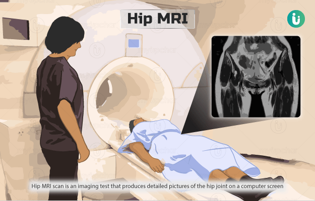
What is a hip MRI? 

Hip MRI (magnetic resonance imaging) scan is an imaging test that produces detailed pictures of the hip joint on a computer screen using a strong magnetic field and radio waves.

The hip joint, one of the largest weight-bearing joints in our body, consists of the ball or rounded head of the thigh bone (femur) cupped into the socket (acetabulum) of the hip bone. The joint is reinforced by tissues such as:

  • Cartilage: The tissue that lines the joint and provides cushioning.
  • Ligaments: Tough bands of tissue that anchor the ball to the socket.
  • Labrum: A circular band of cartilage that surrounds the socket to provide more stability to the joint.

For an MRI procedure, the person is asked to lie under the strong magnets of the MRI machine. The transmitter of the machine sends short bursts of radio waves, which alter the atoms in the part of the body being scanned. Once the transmitter is shut off, the altered atoms send out radio signals, which are picked up by the receiver of the machine. A computer then uses the data received to create images of the scanned body part.

In some cases, contrast materials are administered to the patient to improve the clarity of pictures. These materials are special dyes that outline the structures within the hip joint and help distinguish normal from abnormal conditions. An MRI of joints with contrast, otherwise called MR arthrography, is the preferred technique for the evaluation of joint conditions and unexplained pain

There are two types of MR arthrography: direct MR arthrography and indirect MR arthrography. In direct MR arthrography, the doctor injects the contrast material directly into the joint, whereas in the indirect method, the contrast material is administered into the bloodstream and gets absorbed into the joint. Direct MR arthrography is the preferred method because it enlarges the joint, thereby allowing better visualisation of the internal structures.

  1. Who cannot have a hip MRI?
  2. Why is hip MRI done?
  3. How should I prepare for the hip MRI?
  4. What is the procedure for the hip MRI?
  5. How does a hip MRI feel?
  6. What do the results of a hip MRI scan mean?
  7. What are the risks and benefits of a hip MRI?
  8. What happens after a hip MRI?
  9. What other tests can be done with a hip MRI?
  10. What is the difference between contrast and non-contrast hip MRI?
Doctors for Hip MRI

Your healthcare practitioner may advise against this test in the following situations:

  • Pregnancy: MRI is usually avoided during pregnancy because it is not known if the strong magnetic field induced during the test can have long-term effects on the developing fetus.
  • Tattoos: Some tattoo inks may contain traces of metal, which can cause a burning sensation or discomfort during the scan.
  • Metal inside the body: The strong magnets used for an MRI scan can affect metal objects in the body. MRI cannot be performed without ensuring that the metal is MRI safe.
myUpchar doctors after many years of research have created myUpchar Ayurveda Urjas Capsule by using 100% original and pure herbs of Ayurveda. This ayurvedic medicine has been recommended by our doctors to lakhs of people for sex problems with good results.
Long Time Capsule
₹719  ₹799  10% OFF
BUY NOW

Your healthcare practitioner may order this test if you show any of the following symptoms:

  • Pain or stiffness in the hip
  • Pain in the groin or buttock area
  • Pain in the hip without previous accidents or injury
  • Sharp pain with weight-bearing, which subsides with rest
  • A locking or clicking sound in the hip area with movement
  • Feeling unsteady on standing

You may be asked not to drink or eat anything for four to six hours before the test. Inform the medical staff if you have any of the following:

  • Artificial heart valves
  • Heart defibrillator or pacemaker
  • Kidney diseases or kidney dialysis
  • Inner ear implants
  • Recently placed artificial joints
  • Worked with metal sheets
  • Stents in the blood vessels
  • Clippings placed for a brain aneurysm (bulged weakened blood vessel)

Also, tell your doctor if you have a fear of closed spaces. They may give you medicine that will help you feel sleepy and less anxious.

Since the MRI machine contains strong magnets, you will not be allowed to carry any metal objects (such as hairpins, jewellery, watches, etc.) inside the scan room.

You will be asked to wear a hospital gown or clothes without metal fasteners for the scan.

A hip joint MRI involves the following steps:

  • You will be positioned on a moveable bed that slides into the centre of the tunnel-shaped MRI machine.
  • The technologist may use straps to help you maintain your position and stay still during imaging.
  • A device that would send and receive radio waves will be positioned around your hip joint.
  • If a contrast material is required, it will be administered through either of the following ways:
    • Indirect arthrography:
      • Your doctor will inject the contrast material through an intravenous line in your arm.
    • Direct arthrography:
      • The doctor will first take an X-ray of the hip joint.
      • They will clean the area over the joint with an antiseptic and will inject a numbing medicine (local anaesthetic) into the area.
      • Using ultrasound guidance, the doctor will place a thin needle into your hip joint and inject the contrast material.
      • They may ask you to move the joint to spread the contrast material in the joint.
      • A direct arthrography procedure takes about half an hour.
  • You will then be positioned into the centre of the scanner, and a series of images will be taken.
  • The MRI procedure may take more than an hour to complete.
myUpchar doctors after many years of research have created myUpchar Ayurveda Kesh Art Hair Oil by using 100% original and pure herbs of Ayurveda. This Ayurvedic medicine has been recommended by our doctors to more than 1 lakh people for multiple hair problems (hair fall, gray hair, and dandruff) with good results.
Bhringraj Hair Oil
₹599  ₹850  29% OFF
BUY NOW

During the procedure, the area being scanned may feel slightly warm, which is normal. The machine makes loud sounds when images are being taken, but you will be given earplugs to mask them. You may feel slight discomfort when the contrast material is injected into the vein.

In direct arthrography, you may have a temporary burning sensation when the local anaesthetic is injected. 

You may feel pressure or pain when the contrast is injected into the joint and after the injection, you may feel fullness in the joint and hear gurgling on moving it.

An abnormal result in your MRI scan hip joint could be an indication of any of the following conditions:

  • With contrast
    • Osteonecrosis of the head of the thigh bone (death of bone tissue due to loss of blood supply)
    • Subchondral insufficiency fracture in the head of the thigh bone (thin crack in the bone due to repetitive stress)
    • Transient osteoporosis (temporary bone loss) in the head of the thigh bone
    • Femoroacetabular impingement (friction between the bones that form the hip joint due to growth of extra bone)
    • Acetabular labral tears (injury to the labrum)
    • Perthes disease (a childhood condition in which the blood supply to the head of the thigh bone is temporarily disrupted)
    • Bursitis (swelling of a fluid-filled sac called bursa)
    • Hip synovitis (swelling of the tissues lining the hip joint)
    • Hip arthritis (damage to the cartilage of the hip joint)
    • Hip dislocation (dislocation of the ball of hip joint from the socket)
    • Hip fracture 
    • Osteomyelitis of the hip joint (infection of the joint)
  • Without contrast
    • Tears of ligamentum teres (a ligament within the hip joint)
    • Labral tears (injury to the labrum)
    • Chondral defects (damage to the cartilage)

The risks associated with the MRI test include:

  • The strong magnetic field created by the machine can displace or damage metal implants.
  • The contrast used can worsen kidney diseases in people undergoing dialysis. It can also cause allergic reactions in rare cases.
  • In direct arthrography, there is a risk of infection. There is also a minimal chance of injury to a blood vessel or nerve near the joint.

The benefits of the MRI test include:

  • No radiation exposure, if you are going for an MRI without arthrography
  • A detailed diagnostic picture of the structures within the joint
  • MRI without arthrography is a non-invasive test

(Read more: Best yoga poses for hips)

myUpchar doctors after many years of research have created myUpchar Ayurveda Urjas Energy & Power Capsule by using 100% original and pure herbs of Ayurveda. This Ayurvedic medicine has been recommended by our doctors to lakhs of people for problems like physical and sexual weakness and fatigue, with good results.
Power Capsule For Men
₹719  ₹799  10% OFF
BUY NOW

You can continue with your normal activities soon after completion of the test. However, if you were given a sedative, you need to take the following precautions:

  • Have someone be with you for 24 hours after the test
  • Avoid driving or using public transport for 24 hours
  • Avoid operating any machinery on the day of the test

If the contrast material was used, 

  • You may experience swelling and discomfort in the joint. You can apply ice to lessen the swelling. The symptoms normally disappear after two days. If they still persist, inform your doctor.
  • Avoid any vigorous exercise for at least 24 hours after the test as there is a risk of dislocation of the joint.

(Read more: Exercises to reduce hip pain)

Your healthcare practitioner may order the following tests along with the MRI scan:

  • Local anaesthetic: The medical staff will give you a numbing medicine in the hip joint. If the medicine provides temporary pain relief, it confirms the diagnosis of femoroacetabular impingement.
  • Nuclear medicine bone scan: It shows changes in the bone caused by transient osteoporosis.

Following are a few key differences between contrast and non-contrast hip MRI:

  • A contrast hip MRI is done in certain conditions to improve the clarity of images produced.
  • A contrast hip MRI is usually used to detect conditions like osteonecrosis and subchondral insufficiency fracture in the head of the thigh bone, transient osteoporosis, femoroacetabular impingement, acetabular labral tears and Perthes disease. The conditions detected with a non-contrast hip joint MRI include tears of ligamentum teres, labral tears and chondral defects.
  • A hip MRI with contrast is done in conjunction with direct or indirect arthrography. In a direct arthrography method, the contrast is injected into the joints, while in the indirect method, the contrast is administered intravenously.
  • The risks associated with MRI of the hip with contrast are the same as that of non-contrast hip MRI and include the risks associated with the contrast dye as well. 

(Read more: Hip replacement surgery)

Disclaimer: All results must be clinically correlated with the patient’s complaints to make a complete and accurate diagnosis. The above information is provided from a purely educational point of view and is in no way a substitute for medical advice by a qualified doctor.

Dr. Rachita Gupta

Dr. Rachita Gupta

Radiology
12 Years of Experience

Dr. Tejinder Kataria

Dr. Tejinder Kataria

Radiology
35 Years of Experience

Dr. Shyam Singh Bisht

Dr. Shyam Singh Bisht

Radiology
17 Years of Experience

Dr. Shikha Goyal

Dr. Shikha Goyal

Radiology
18 Years of Experience

Read on app
cross
Ask your health query from live doctors now!