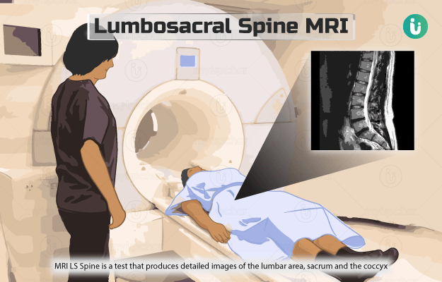
What is a Lumbosacral (LS) spine MRI? 

Magnetic resonance imaging lumbosacral spine (MRI LS Spine) is a test that produces detailed images of the lumbar area (lower part of the spine), sacrum (base of the spine) and the coccyx (the tailbone). The test helps to identify or look for any problems in these areas of the spine.

Unlike an x-ray and CT scan machine, the MRI machine does not use any radiation. Instead, it uses a large magnet, radio waves and a computer for producing images of the internal structures of the body. During the scan, the magnets and radio waves align and realign the protons (parts of atoms) of the body in a particular way. This whole process is read by an attached computer, which uses the data to create images of the scanned area. 

An MRI is sometimes done with a contrast dye, injected into the patient’s body right before the scan, The dye binds with body tissues and helps provide clearer images.

  1. Why is a lumbosacral (LS) spine MRI done?
  2. Who cannot have a lumbosacral (LS) spine MRi?
  3. How should I prepare for a lumbosacral (LS) spine MRI?
  4. How is a lumbosacral (LS) spine MRI done?
  5. How will a lumbosacral (LS) spine MRI feel like?
  6. What do the results of a lumbosacral (LS) spine MRI mean?
  7. What are the risks and benefits of a lumbosacral (LS) spine MRI?
  8. What happens after a lumbosacral (LS) spine MRI?
  9. What are the other tests that can be done with a lumbosacral (LS) spine MRI?
Doctors for Lumbosacral spine MRI

Your healthcare practitioner may order this test if you have:

MRI LS scan with a contrast dye is especially used in the following conditions:

  • To differentiate causes of lower back pain and lumbar radiculopathy
  • To distinguish recurrent disc herniation after operation of spine
  • To show nerve roots in viral or inflammatory conditions
  • To detect tumours
myUpchar doctors after many years of research have created myUpchar Ayurveda Urjas Capsule by using 100% original and pure herbs of Ayurveda. This ayurvedic medicine has been recommended by our doctors to lakhs of people for sex problems with good results.
Long Time Capsule
₹719  ₹799  10% OFF
BUY NOW

This test is very safe for most people. However, it may not be recommended in the following conditions:

  • If you have metal implants or fragments: It does not necessarily mean you cannot undergo the test. The final decision will be taken by the medical staff carrying out the scan. In some cases, it is possible to make metal implants, like pacemakers, MRI-safe.
  • If you have tattoos on your body: Most tattoos are safe, but some tattoo ink may contain traces of metal. Inform the radiography staff if you feel heat or discomfort in your tattoo during the scan.

Generally, you are not supposed to eat or drink for four to six hours before the scheduled time of the procedure. If you have a fear of closed spaces (claustrophobia), you may be given a sedative or anaesthesia that will help you feel sleepy or less anxious. 

Also, tell your healthcare provider or radiologist if you have:

  • Brain aneurysm clips
  • Pacemaker or heart defibrillator
  • Artificial heart valves
  • Cochlear (inner ear) implant
  • Artificial joints that have been placed recently
  • Vascular stents
  • Kidney disease/dialysis
  • Worked with sheet metal in the past

An MRI scan usually involves the following steps:

  • The radiography staff will ask you to remove all metal objects, such as a watch, jewellery and keys, before entering the scan room.
  • They may give you a hospital gown to change into.
  • You will have to lie on the scanner’s narrow table. The table will slide into the large cylindrical tube of the MRI machine. 
  • If the procedure requires a contrast dye, it may be administered into your body through IV or injection before the test. The contrast helps to view certain areas more clearly.
  • The test may last for 30-60 minutes or longer.
myUpchar doctors after many years of research have created myUpchar Ayurveda Kesh Art Hair Oil by using 100% original and pure herbs of Ayurveda. This Ayurvedic medicine has been recommended by our doctors to more than 1 lakh people for multiple hair problems (hair fall, gray hair, and dandruff) with good results.
Bhringraj Hair Oil
₹599  ₹850  29% OFF
BUY NOW

MRI is a painless procedure. You might feel the table of the MRI scanner to be hard/cold. In such instances, you can ask for a pillow or blanket. The scanner may make loud noises during the procedure. The radiography staff will provide you with earplugs to help reduce the noise.

The MRI images will be interpreted by the radiologist, who will send a detailed report to your physician. Then, the physician will discuss the results with you. Abnormal results may be due to any of the following conditions:

  • Spinal stenosis (narrowing of the spinal column)
  • Lumbar radiculopathy (herniated or slipped disc)
  • Spondylitis (abnormal wearing of the spinal bones and cartilage)
  • Degenerative changes due to age
  • Bone infection
  • Osteoporosis resulting in fracture of the lower back
  • Spinal cord injury/abscess
  • Syringomyelia (a disorder characterised by a fluid-filled pocket formed within the spinal cord)
  • Ankylosing spondylitis (inflammation of the joints that primarily affects the spine)
  • Cauda equine syndrome (damage to a group of nerves that spread out from the bottom of the spinal cord)
  • Diskitis (inflammation of the disk)
    • Spinal tumour
    • Infection of the sacroiliac joint (joint between the sacrum and a hip bone)
    • Sacral meningeal cysts (fluid-filled sacs that affect the nerves of the sacral region)
    • Sacral fractures
    • Sacral fatigue stress fracture (due to abnormal stresses from intense training
    • Sacral tumours
    • Sacral agenesis observed in children (absence of the sacrum and coccyx and portions of lumbar spine)

 The test involves a safe scanning procedure. Some complications that may occur are as follows:

  • In rare cases, the contrast injected before the test may cause an allergic reaction.
  • The scanner can disrupt the working of pacemakers and other implants.
myUpchar doctors after many years of research have created myUpchar Ayurveda Urjas Energy & Power Capsule by using 100% original and pure herbs of Ayurveda. This Ayurvedic medicine has been recommended by our doctors to lakhs of people for problems like physical and sexual weakness and fatigue, with good results.
Power Capsule For Men
₹719  ₹799  10% OFF
BUY NOW

If sedatives were taken for the test, you might need to rest until its effects wear off and avoid driving or using heavy machinery. 

If you were injected with contrast during the procedure, you would be monitored for side effects like swelling, itching, rash and difficulty in breathing. In case you see any redness or swelling at the site of IV or injection, tell your healthcare practitioner as it may be a sign of infection.

Some of the additional tests that can be performed along with this test include:

Disclaimer: All results must be clinically correlated with the patient’s complaints to make a complete and accurate diagnosis. The above information is provided from a purely educational point of view and is in no way a substitute for medical advice by a qualified doctor.

Dr. Rachita Gupta

Dr. Rachita Gupta

Radiology
12 Years of Experience

Dr. Tejinder Kataria

Dr. Tejinder Kataria

Radiology
35 Years of Experience

Dr. Shyam Singh Bisht

Dr. Shyam Singh Bisht

Radiology
17 Years of Experience

Dr. Shikha Goyal

Dr. Shikha Goyal

Radiology
18 Years of Experience

References

  1. Curry BP, Rosner MK. Evaluation and treatment of lumbar disk disease. In: Winn HR, ed. Youmans and Winn Neurological Surgery. 7th ed. Philadelphia, PA: Elsevier; 2017:chap 286
  2. Miller MD, Thompson SR, eds. DeLee and Drez's Orthopaedic Sports Medicine. 5th ed. Philadelphia, PA: Elsevier Saunders. 2018.
  3. Chou R, Qaseem A, Owens DK, Shekelle P; Clinical Guidelines Committee of the American College of Physicians. Diagnostic imaging for low back pain: advice for high-value health care from the American College of Physicians. Ann Intern Med. 2011;154(3):181-189. PMID: 21282698
  4. Gardocki RJ, Park AL. Degenerative disorders of the thoracic and lumbar spine. In: Azar FM, Beaty JH, Canale ST, eds. Campbell's Operative Orthopaedics. 13th ed. Philadelphia, PA: Elsevier; 2017:chap 39
  5. Wilkinson ID, Graves MJ. Magnetic resonance imaging. In: Adam A, Dixon AK, Gillard JH, Schaefer-Prokop CM, eds. Grainger & Allison's Diagnostic Radiology: A Textbook of Medical Imaging. 6th ed. Philadelphia, PA: Elsevier Churchill Livingstone; 2015:chap 5
  6. Nemours Children’s Health System [Internet]. Jacksonville (FL): The Nemours Foundation; c2017; Magnetic Resonance Imaging (MRI): Lumbar Spine
  7. Cleveland Clinic [Internet]. Ohio. US; Magnetic Resonance Imaging (MRI)
  8. National Health Service [Internet]. UK; MRI scan
  9. National Center for Complementary and Integrative Health [Internet]. National Institute of Health. US Department of Health and Human Services; Multiple Sclerosis
  10. Better health channel. Department of Health and Human Services [internet]. State government of Victoria. Australia; MRI scan
  11. National Institute of Neurological Disorders and Stroke [Internet[. National Institute of Health. US Department of Health and Human Services. US; Syringomyelia Information Page
  12. MedlinePlus Medical Encyclopedia [Internet]. US National Library of Medicine. Bethesda. Maryland. USA; Ankylosing spondylitis
  13. Genetics and Rare Diseases Information Center [internet]. National Center for Advancing Translational Sciences. National Institute of Health: US National Library of Medicine. US Department of Health and Human Services; Cauda equina syndrome
  14. Diel J, Ortiz O, Losada RA, Price DB, Hayt MW, Katz DS. The sacrum: pathologic spectrum, multimodality imaging, and subspecialty approach. Radiographics. 2001 Jan-Feb; 21(1): 83-104. PMID: 11158646
  15. Raj MA, Ampat G, Varacallo M. Sacroiliac Joint Pain. [Updated 2021 Jan 29]. In: StatPearls [Internet]. Treasure Island (FL): StatPearls Publishing; 2021 Jan
  16. Figueroa C, Le PH. Anatomy, Bony Pelvis and Lower Limb, Pelvis Bones. [Updated 2020 Sep 16]. In: StatPearls [Internet]. Treasure Island (FL): StatPearls Publishing; 2021 Jan
  17. Yoder K, Bartsokas J, Averell K, McBride E, Long C, Cook C. Risk factors associated with sacral stress fractures: a systematic review. J Man Manip Ther. 2015 May; 23(2): 84-92. PMID: 26109829.
  18. Johns Hopkins Medicine [Internet]. The Johns Hopkins University, The Johns Hopkins Hospital, and Johns Hopkins Health System; Magnetic Resonance Imaging (MRI) of the Spine and Brain
  19. Amukotuwa SA, Cook MJ. Spinal disease: neoplastic, degenerative, and infective spinal cord diseases and spinal cord compression. Neurology and Clinical Neuroscience. 1999; 511-38.
  20. Bradley WG. Use of contrast in MR imaging of the lumbar spine. Magn Reson Imaging Clin N Am. 1999; 7(3): 439–vii. PMID: 10494528.

Related Articles

Upper GI Endoscopy

Dr. Ayush Pandey
MBBS,PG Diploma
7 Years of Experience

Obesity Profile

Dr. Ayush Pandey
MBBS,PG Diploma
7 Years of Experience

Extractable Nuclear Antigen ...

Dr. Ayush Pandey
MBBS,PG Diploma
7 Years of Experience
Read on app
cross
Ask your health query from live doctors now!