Arthrogryposis

Dr. Suvansh Raj NirulaMBBS

February 13, 2021

October 14, 2021

Arthrogryposis
Arthrogryposis

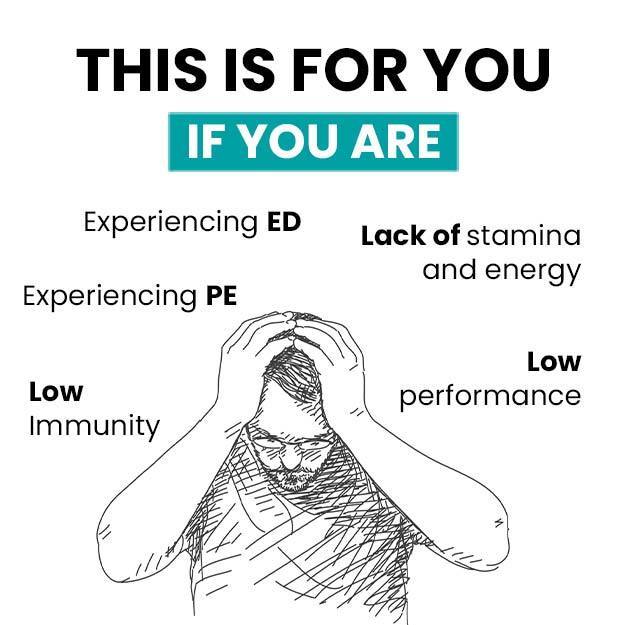
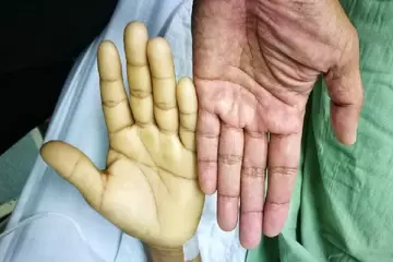
Arthrogryposis, or arthrogryposis multiplex congenita (AMC), is a group of disorders in which multiple (two or more) joint contractures (tightening of normally elastic tissue) are present at birth. Muscle tissue is fibrosed due to lack of mobility during gestational development in pregnancy. Broadly, three categories of arthrogryposis exist – amyoplasia, distal and syndromic arthrogryposis. Genetic and environmental factors can produce arthrogryposis. Certain factors affecting the pregnancy, childbirth and delivery may also be involved. In case of family history of the disease, genetic testing should be offered to expecting parents. Lower limbs are more affected than upper limbs. A diagnosis is made by taking a thorough medical history, clinical examination, radiological imaging and cytogenetic investigations. AMC would need to be managed by a health team led by a paediatrician and a mix of physiotherapy, serial splinting and casting, surgical correction of soft tissue deformities and contractures and psychological care. As the disease is not progressive, unless caused in conjunction with other more serious neurogenic causes, the prognosis is generally favourable.

Types of arthrogryposis

Arthrogryposis multiplex congenita (AMC), is the term used to describe various disorders in which multiple (two or more) non-progressive joint contractures are present at birth. Although joints may develop normally in early pregnancy, the condition can develop due to reduced and restricted movements in the latter half of pregnancy because of connective tissue or neurological disorders. The lack of joint mobility during development can cause most of the muscle tissue to be replaced by fat or fibrous tissue. This muscle fibrosis produces contractures. Following are the three main groups of arthrogryposis multiplex congenita:

  • Amyoplasia: Amyoplasia translates to no muscle growth and is marked by severe muscle weakness and joint contractures. Certain presenting characteristics of amyoplasia in the upper limb include internally rotated shoulders, with extended elbows and flexed wrists that are deviated towards the body. In the lower limbs, dislocated hip, extended knees and severe equinovarus deformities (secondary clubfoot) are common.
  • Distal arthrogryposis: A group of autosomal dominant genetically inherited disorders in which multiple (two or more) joint contractures are present, only in the hands and feet (i.e. in the distal limbs) and are unrelated to any primary neurological or muscular disease. Ten distinct types of distal arthrogryposis have been described.
  • Syndromic arthrogryposis: Joint contractures caused secondary to a primary neurological or muscular disorder are placed under the category of syndromic arthrogryposis. Of these, neurological causes are the most common and important and are mainly of two types – central neurological and neuromuscular.
    • Central neurological: Motor neurone disease affecting the spinal, facial and brainstem motor neurons can result in decreased mobility of the developing foetus and produce arthrogryposis. Spinal muscular atrophies can also cause arthrogryposis. Other causes could be damage to the forebrain. Hyperactive reflexes, unilateral arthrogryposis or cognitive deficits are present on examination and can be anatomically localized by magnetic resonance imaging (MRI) of the brain.
    • Neuromuscular: Genetically linked congenital myopathy and congenital neuropathy are important causes. Genetic peripheral neuropathy is a very rare cause of syndromic arthrogryposis. Electromyography is a very useful investigation to study deficits in muscle and nerve impulse conduction that can explain muscle weakness.
myUpchar doctors after many years of research have created myUpchar Ayurveda Urjas Capsule by using 100% original and pure herbs of Ayurveda. This ayurvedic medicine has been recommended by our doctors to lakhs of people for sex problems with good results.
Long Time Capsule
₹719  ₹799  10% OFF
BUY NOW

Signs and symptoms of arthrogryposis

Amyoplasia presents with signs in the upper and lower limbs; however, the lower limbs are more predominantly affected.

  • Upper limbs:
    • Shoulders – rotated inward and glued close to the body (internally rotated and adducted)
    • Elbows – either extended or fixed in a flexed position
    • Wrists – are deviated toward the body (ulnar deviation)
    • Hands – deformity of thumb and palm and rigid interphalangeal joints (finger joints)
  • Lower limbs: 
    • Hips – one or both of the sides are dislocated
    • Knees – fixed in either an extended or bent position
    • Feet – rigid bilateral clubfeet (secondary talipes equinovarus)
  • Other deformities of the limbs: 
    • Shortening of limbs (especially legs)
    • Webs
    • Dimples
    • Compression due to cord wrapping
    • Absent patellae (kneecaps)
    • Dislocated radial (radius bone of forearm) head
  • Deformities of face and jaw:
    • Asymmetry
    • Flat nasal bridge
    • Haemangiomata
    • Micrognathia
    • Trismus (lock jaw)
  • Other signs and symptoms:
    • Thin subcutaneous tissue and absent skin creases
    • Symmetrical deformities that are more severe distally
    • Rigid joints
    • Congenital dislocation of the hips (and sometimes the knees)
    • Atrophy or absence of groups of muscles
    • Contractures (especially of distal joints)
    • Pterygia (these are winglike triangular membranes occurring typically in the neck, knees, elbows, ankles or fingers)
    • Scoliosis
    • Genital deformity
    • Umbilical hernias
    • Inguinal hernias
    • There may be many other malformations of the skeleton, respiratory tract, urinary system and nervous system

(Get online doctor consultation for any health issue)

Causes of arthrogryposis

Arthrogryposis results from restriction of the mobility of the fetus, which hampers the growth of muscles. The causes of this restriction can be:

  • Environmental: 
    • Maternal oligohydramnios (reduced amniotic fluid, in which the developing baby is suspended inside the womb during pregnancy can cause the limbs to be compressed abnormally against the walls of the uterus)
    • Maternal infections during pregnancy:
    • Fetal akinesia
    • Maternal myasthenia gravis (a very rare cause)
  • Genetic:
    • X-linked recessive
    • Autosomal dominant
    • Autosomal recessive
    • Mitochondrial
    • Chromosomal abnormalities like trisomy of chromosome 18
  • Musculopathy:
    • Congenital myopathies
    • Congenital muscular dystrophy
    • Myasthenic syndromes
    • Intrauterine viral myositis
    • Mitochondrial disorders
  • Connective tissue disorders:
    • Diastrophic dysplasia
    • Osteochondroplasia
    • Metatropic dwarfism
  • Neurogenic disorders: Neurological abnormalities are responsible for the most severe forms of arthrogryposis. Examples include:
    • Myelomeningocele
    • Sacral agenesis
    • Spinal muscular atrophy (anterior horn cell disease of prenatal origin SMA 0, not Werdnig-Hoffman SMA 1)
    • Congenital contracture syndrome
    • Cerebro-oculo-facial syndrome
    • Marden-Walker syndrome
    • Pena-Shokeir syndrome

Risk factors of arthrogryposis

Following are some of the risk factors for the development of arthrogryposis:

  • Pregnancy factors: 
    • Oligohydramnios (as in Potter's syndrome)
    • Multiple births
  • Maternal infections:
  • Maternal Drug intake (both therapeutic and recreational):
  • Maternal trauma
  • Uterus abnormalities: A septate or bicornuate uterus can reduce the room for movement available to the growing baby
  • Intrauterine vascular abnormalities/compromise
  • Other maternal illnesses:
myUpchar doctors after many years of research have created myUpchar Ayurveda Kesh Art Hair Oil by using 100% original and pure herbs of Ayurveda. This Ayurvedic medicine has been recommended by our doctors to more than 1 lakh people for multiple hair problems (hair fall, gray hair, and dandruff) with good results.
Bhringraj Hair Oil
₹599  ₹850  29% OFF
BUY NOW

Diagnosis of arthrogryposis

The doctor begins by taking a proper medical history with a special focus on family history and history about the pregnancy, childbirth and delivery.

Family history:

  • Other affected children or members of the family
  • Consanguinity (sexual relations between people who share an ancestor), as it increases the risk of rare recessive disorders
  • Advanced parental age (both maternal and paternal) increases the overall risk for chromosomal disorders
  • A parent who may have suffered from a mild form of arthrogryposis in childhood or had experienced infantile contractures
  • A history of miscarriages due to possible fetal abnormalities caused by genetic disorders
  • Diseases like myasthenia gravis, multiple sclerosis (MS) and myotonic dystrophy in the mother can be a rare cause of arthrogryposis in the baby

Pregnancy, childbirth and delivery related history:

  • Infections suffered by the mother during pregnancy caused by some viruses, like rubella and Coxsackie, can cause neuropathy in the baby
  • Prolonged maternal pyrexia (raised body temperature, due to fever or hot saunas or baths used in the childbirth process) can produce contractures due to abnormal nerve growth and migration.
  • Drugs, both medicinal (like phenytoin) and recreational (alcohol), taken while pregnant can impair foetal movements and precipitate arthrogryposis.
  • Oligohydramnios (reduced amniotic fluid) during pregnancy reduces fetal movements.
  • Abnormalities of uterus structure like septate uterus or large fibroids can compress the foetus and prevent mobility.
  • An abnormal lie or a complicated delivery due to an unusual position of the baby during childbirth (like breech, where the feet come first) can cause problems.
  • Abnormalities of the placenta, for example, a short umbilical cord can get wrapped around the baby’s limbs and inhibit movements.
  • Multiple pregnancies (like twins, triplets, etc.) can also restrict movement in the womb.

After taking a thorough medical history, the specialist will conduct a physical examination. After inspection of the characteristic features as described above, a total systemic examination will also be done. Special attention will be given to neurological testing as this can help differentiate syndromic arthrogryposis caused secondary to neurogenic disorders from other causes. Classifying arthrogryposis is crucial to correct diagnosis, effective treatment and the prognosis.

Tests for arthrogryposis

Following are the investigations that may be required for arthrogryposis:

  • Genetic testing: In case of a history of arthrogryposis in the family, genetic counselling and testing should be sought out.
  • Blood test: Serum creatinine kinase can diagnose a muscular disorder especially in the case of obvious muscular deformity.
  • Radiological imaging:
    • X-rays: Of deformed joints and bones
    • Ultrasound: Of muscle groups in the deformed body parts
    • CT scans: Both of the deformities and the central nervous system (CNS) structures (brain and spinal cord) to diagnose possible neurological causes.
    • MRI: Another modality that can be used for both deformities and the brain and spinal cord.
  • Cytogenetic testing: Testing for genetic and chromosomal abnormalities can elucidate the true underlying cause of arthrogryposis.

Treatment of arthrogryposis

Arthrogryposis can be managed through an integrated approach with physiotherapy, splinting, casting, surgery (where necessary) and psychosocial care.

Conservative management: Conservative management is useful, particularly in cases due to amyoplasia and distal arthrogryposis. However, it may not be very beneficial in syndromic arthrogryposis due to more severe neurogenic and myopathic disorders.

  • Physiotherapy: Stretching exercises to improve mobility, prevent further contractures and improve the range of motion of joints should be started as early as possible.
  • Splinting: In between physical therapy sessions, splinting helps correct and maintain the deformed joints being treated.
  • Casting: Weekly changing of casts following manual manipulation (holding, stretching and moving) of the affected joints is also useful. Techniques using this principle include the Ponseti method used specifically for clubfeet.

Surgical management: Surgery can sometimes be necessary to correct soft tissue contractures and joint deformities. Operative management is often done for the following reasons:

  • To stabilize dislocated hips
  • To stabilize spinal deformities
  • For wrists and thumb deformities
  • Clubfeet, etc.

However, patients can often be at risk of developing malignant hyperthermia and thus anaesthesia (putting the patient under sedation for surgery) can be challenging.

Psychological care: The extent of psychological care needed varies based on the extent of the disease, the patient’s mental health and other social circumstances. It is important to not overlook it. Learning to lead meaningful lives with this restricting disease is essential to the overall health of the patient.

Occupational therapy: In extreme cases where normal physical and physiological functioning is impaired, occupational therapy can help lead productive and normal lives.

myUpchar doctors after many years of research have created myUpchar Ayurveda Urjas Energy & Power Capsule by using 100% original and pure herbs of Ayurveda. This Ayurvedic medicine has been recommended by our doctors to lakhs of people for problems like physical and sexual weakness and fatigue, with good results.
Power Capsule For Men
₹719  ₹799  10% OFF
BUY NOW

Prognosis of arthrogryposis

Prognostic of the disease depends on the underlying cause of arthrogryposis; however, most patients have a normal lifespan. Patients with amyoplasia tend to have a better outcome. Scoliosis appears in most cases at some point and should be corrected before it becomes too severe to improve the outlook.

Prevention of arthrogryposis

In the case of prior family history of arthrogryposis or other associated disorders, genetic counselling and testing should be sought by expecting parents. Maternal infections, unsupervised drug intake, pyrexia and trauma during pregnancy are modifiable risk factors that can be avoided to prevent certain kinds of arthrogryposis.


References

  1. Bamshad M, Van Heest AE, Pleasure D. Arthrogryposis: A Review and Update. J Bone Joint Surg Am. 2009 Jul 1; 91(Suppl 4): 40–46. PMID: 19571066.
  2. Ma L, Yu X. Arthrogryposis multiplex congenita: classification, diagnosis, perioperative care, and anesthesia. Front Med. 2017 Mar;11(1):48-52. PMID: 28213879.
  3. Ayadi K, Trigui M, Abid A, Cheniour A, Zribi M, Keskes H. Arthrogryposis: Clinical manifestations and management. Arch Pediatr. 2015 Aug;22(8):830-9. PMID: 26141802.
  4. Hamdy RC, et al. Treatment and outcomes of arthrogryposis in the lower extremity. Am J Med Genet C Semin Med Genet. 2019 Sep;181(3):372-384. PMID: 31479584.
  5. O'Flaherty P. Arthrogryposis multiplex congenita. Neonatal Netw. 2001 Jun;20(4):13-20. PMID: 12143898.
  6. Hall J G. Arthrogryposis multiplex congenita: etiology, genetics, classification, diagnostic approach, and general aspects. J Pediatr Orthop B. 1997 Jul;6(3):159-66. PMID: 9260643.