Clubfoot

Dr. Suvansh Raj NirulaMBBS

January 23, 2021

January 27, 2021

Clubfoot
Clubfoot

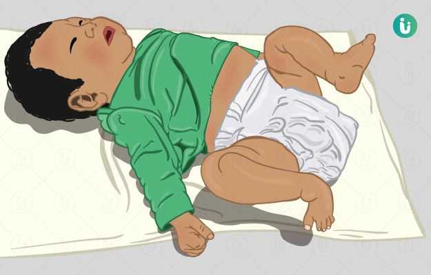
Clubfoot, or congenital talipes equinovarus, is a foot deformity in which the foot is rotated from its normal position, downward and inward. It is a relatively common birth defect and while it is usually diagnosed on examination of the neonate, it can also be diagnosed by ultrasound during pregnancy. Although result of a mix of genetic and environmental factors, some modifiable maternal risk factors like smoking during pregnancy and maternal obesity can also play a part. Treatment of clubfoot is generally conservative. The standard method employed, Ponseti method, involves serial manual manipulation (holding, stretching and moving) the baby’s foot into the correct position and shape and then weekly application of a foot to groin cast for several months. After a natural shape and position are achieved, the Achilles’ tendon is lengthened and the foot is placed in specialised braces and shoes for a few months. Surgical correction can be needed in cases that fail to respond to conservative management for over a year or have residual undercorrected deformities following it. 

(Read more: Pregnancy week by week)

Types of clubfoot

Clubfoot refers to a characteristic foot deformity. Primary idiopathic clubfoot, also known as congenital talipes equinovarus (CTEV), is an orthopaedic birth defect of one or both feet. The affected foot is rotated (talipes) from its normal position downward (equinus, like a horse’s hoof) and inward (varus). In about half of the cases, both feet are affected. Clubfoot has an incidence of around 1 to 4 per 1,000 births. Furthermore, most of these cases arise in resource-poor developing countries with limited access to healthcare. If one twin is affected, there is a one in three chance of the other twin having a clubfoot as well. A mix of genetic and environmental causes are to blame for giving rise to talipes equinovarus, however, the underlying defect remains the same – undue shortening of the Achilles’ tendon (which attaches the calf muscles to the ankle joint) – and causes the abnormally rotated position of the foot. There are three types of clubfoot:

  • Primary Idiopathic clubfoot: CTEV arises due to idiopathic (unknown) causes and is twice as likely to present in male babies at birth. 
  • Neurogenic clubfoot: This type of clubfoot is usually associated with another neurological birth defect, for example, spina bifida (a neural tube defect in which the spine and spinal cord do not form properly, often resulting in an outpouching of membranes at the back called meningocele or myelomeningocele). Other neurological conditions can give rise to the development of clubfoot later in childhood. Causes of neurogenic clubfoot include:
  • Syndromic clubfoot: Clubfoot can arise along with a myriad of other clinical signs and symptoms that form part of various syndromes. Examples include:
    • Arthrogryposis
    • Constriction band syndrome
    • Tibial hemimelia
    • Diastrophic dwarfism

(Read more: Achilles tendon problems)

myUpchar doctors after many years of research have created myUpchar Ayurveda Urjas Capsule by using 100% original and pure herbs of Ayurveda. This ayurvedic medicine has been recommended by our doctors to lakhs of people for sex problems with good results.
Long Time Capsule
₹719  ₹799  10% OFF
BUY NOW

Signs and symptoms of clubfoot

The presentation of clubfoot may involve:

  • The front or top portion of the foot is rotated downward and inward, increasing the arch and turning the heel inward
  • The overall length of the affected leg is reduced
  • Calf muscle bulk is lesser in the affected leg 
  • Abnormal creases on the foot 

(Read more: Habits to avoid to ensure a safe and healthy pregnancy

Despite its appearance, clubfoot is generally painless, especially if the child hasn’t started walking yet. 

Causes of clubfoot

It is not fully understood how clubfoot occurs and the deformity is said to be idiopathic. However, genetics, environment and a combination of both are said to be involved. The main underlying defect, irrespective of the cause, remains the same – shortening and tightening of the Achilles’ tendon, which joins the calf muscles to the ankle joint, and the subsequent shortening of the ligaments of the foot bones that give it an abnormal shape. Some hypotheses to explain CTEV have been presented by researchers: 

  • Underdevelopment of bones and muscles in the embryonic foot.
  • Connective tissue abnormalities that cause an increase in fibrous tissue and cause muscle contractures, tightening of tendons and shortening of ligaments. (Read more: Connective tissue disease
  • Amniocentesis during pregnancy for genetic testing can cause amniotic fluid to leak out. This reduction in fluid volume can cause the baby’s feet, which are normally suspended in the liquor amnii, to become compressed against the walls of the mother’s womb and grow abnormally. 
  • Genetic disorders involving the genes coding for the muscle contractile proteins.
  • Maternal smoking during pregnancy has been identified as a modifiable risk factor. 
  • Maternal obesity is another modifiable risk factor significantly associated with clubfoot. 
  • Selective Serotonin Reuptake Inhibitors (SSRIs) are drugs used for the treatment of depression and other associated mental health disorders. Some studies suggest that the exposure of the fetus to SSRI antidepressants being taken by the mother can increase the risk for clubfoot to some extent. (Read more: Depression during pregnancy)
  • It can be secondary to other conditions, most notably distal arthrogryposis and meningocele. 

(Read more: Mental Illness

Risk factors for clubfoot

Following are some risk factors for clubfoot: 

  • Gender, as males are twice as likely to have clubfoot than females 
  • Family history of clubfoot, such as a parent or sibling with the condition
  • Smoking during pregnancy
  • Using recreational drugs during pregnancy
  • Too little amniotic fluid surrounding the baby during pregnancy, which is oligohydramnios
  • Other skeletal birth defects
myUpchar doctors after many years of research have created myUpchar Ayurveda Kesh Art Hair Oil by using 100% original and pure herbs of Ayurveda. This Ayurvedic medicine has been recommended by our doctors to more than 1 lakh people for multiple hair problems (hair fall, gray hair, and dandruff) with good results.
Bhringraj Hair Oil
₹599  ₹850  29% OFF
BUY NOW

Diagnosis of clubfoot

History: A maternal family history of clubfoot or a personal history of smoking during pregnancy is taken. Clubfoot may also be detected ultrasonographically on the scans, especially with a level 2 ultrasound, done during pregnancy. 

(Read more: How to quit smoking)

Neonatal examination: All newborn babies are examined from head to toe upon birth for possible birth defects. Clubfoot is usually diagnosed upon this neonatal examination. (Read more: Newborn health care tips

  • Four components of clubfoot deformity: 
    • Cavus: A high-arched foot with a “cave” like appearance. 
    • Adductus: The front portion of the foot is curved inwards towards the big toe.
    • Varus: The heel of the foot is turned in causing the child to place their foot on its side while walking
    • Equinus: Due to the short and tight Achilles’ tendon, the foot is forced to point downward in a fixed tiptoe position. As this resembles the hoof of a horse, it is called 'equinus'. 
  • Factors measuring the severity of deformity:
    • Stiffness of the deformity (estimating the extent to which it can be corrected by manual manipulation of the foot)
    • Presence of skin creases at the arch and heel
    • Poor muscle consistency
  • Pirani score: Is a simple visual scoring system used by orthopaedic surgeons to determine the severity and monitor the patient’s progress for assessment and treatment of clubfoot. Scores range from 0 to 6. Higher the score allotted, greater the severity. 

(Read more: Check up during pregnancy)

Tests for clubfoot

Although clubfoot is usually a clinical diagnosis, it can be diagnosed before birth by an obstetric ultrasound. Level two ultrasound, done between 18 to 24 weeks of pregnancy, is most useful for this purpose. In mild cases of clubfoot, when the clinical diagnosis is dubious, an X-ray of the foot might occasionally be ordered by the doctor. 

Management of clubfoot

In older times, major surgery was needed to correct clubfoot. However, if the treatment is started on time, surgery is seldom needed now. Three methods of conservative management exist – Ponseti method (the most common and preferred), French method (also used clinically today but is less superior to the Ponseti method) and Kite (an older technique which is rarely used now). However, additional surgery after conservative treatment to correct clubfoot may be needed in 20% of the cases.

Conservative: 

  • Ponseti Method: Ponseti method is the standard treatment for clubfoot. Broadly it involves the following steps:
    • Repositioning the baby’s deformed foot: By gentle manual manipulation (holding, stretching and moving), the orthopedician moulds the baby’s foot into a normal shape and position. This does not cause the baby any pain or discomfort. 
    • Serial casting: After holding the baby’s affected foot in the correct position, a special cast is applied that extends from the baby’s foot to the groin. This cast is changed once a week for several months. 
    • Achilles’ tendon lengthening: After several months of serial foot manipulation and casting, a minor surgical release of the Achilles’ tendon is made to loosen it. This is called a percutaneous Achilles tenotomy. It is done by making an incision on the tendon, at the back of the foot near the ankle where it meets the joint. It is done under a local anaesthetic. After releasing the tendon, the foot is put in a cast one final time for around three weeks.
    • Stretching exercise: After the clubfoot has been corrected, parents are asked to do regular foot stretching exercises with their baby to prevent relapse. 
    • Bracing: After the clubfoot has been corrected, to prevent any relapses, special braces have to be applied on the baby’s feet and legs. At first, they are to be worn full time for a few months, followed by only at night and lastly only during naps. Various braces exist that can be prescribed for this purpose, the most common one being the Dennis Brown splint which looks like a pair of strap-on boots connected by a bar at the soles. The aim is to correct the clubfoot before the child begins school to avoid the use of braces then. 
    • Special shoes: Similar to leg and foot braces, shoes with a specially designed sole are made for the child. They are to be worn regularly for a few months to a few years to prevent relapse. 
  • French Method: Also known as the functional method, it utilises physiotherapy to correct clubfoot. At first, physical therapy is administered daily for two months, followed by thrice a week sessions for the next four months. In each session, the baby’s feet are manipulated, stretched and taped. After this, home exercises and bracing are carried out to maintain the correction of clubfoot. Although it allows the baby more mobility in the absence of weekly serial casting, it has a lower success rate compared to the Ponseti method due to improper compliance. It can be used in conjunction with the Ponseti method as well. 

Surgical: 

In cases where conservative management of clubfoot has been tried for over a year and failed to produce results, surgical management may be necessary. The basic aim of surgical correction is to lengthen the ligaments and fascia of the foot to correct the components of the clubfoot deformity (cavus, adductus, varus and/or equinus). The operation done to correct all four components in a typical clubfoot is called the Posteromedial Soft Tissue Release (PMSTR). The foot bones are exposed surgically and the ligaments and fascia of the foot are released sequentially till the foot achieves a normal position and shape. Wires will be put to hold the new position of foot bones and will be removed in a few weeks followed by casting for a few months. This operation is done at 9 to 12 months of age. 

For undercorrected clubfoot following conservative management, soft tissue release operations can be done in accordance with the component of clubfoot deformity that needs correction. 

myUpchar doctors after many years of research have created myUpchar Ayurveda Urjas Energy & Power Capsule by using 100% original and pure herbs of Ayurveda. This Ayurvedic medicine has been recommended by our doctors to lakhs of people for problems like physical and sexual weakness and fatigue, with good results.
Power Capsule For Men
₹719  ₹799  10% OFF
BUY NOW

Complications of untreated clubfoot

Complications or problems associated with clubfoot generally arise only when the baby begins to stand and walk. Difficulties with the following can be experienced then:

  • Movement: Flexibility of the affected foot is slightly reduced in comparison. (Read more: How to improve flexibility
  • Leg length: The overall length of the leg with the affected foot may be reduced. However, mobility isn’t affected as such.
  • Shoe size: Due to the abnormal shape, the affected foot can be up to 1½ shoe sizes smaller compared to the unaffected foot. 
  • Calf size: Owing to the abnormally shortened and contractured muscles and tendons of the leg with clubfoot, the calf on the affected side can be smaller. 

Untreated clubfoot can lead to:

  • Arthritis: Abnormal weight bearing due to the deformity can result in wear and tear of bone surfaces of joints, producing arthritis in these children. 
  • Calluses and sores on foot: The skin on the surface of the foot in contact with the floor while walking abnormally can lead to the development of calluses and sores. This is due to the fact that the skin in contact with the ground in clubfoot is not as thick and hardy as that of the sole which is designed to naturally touch the floor while walking. 
  • Inability to walk normally: As the ankle joint is rotated from its usual position, the child is unable to place the sole on the ground while walking and the deformed foot is placed on the floor in order to pace forward in an abnormal manner. This results in an unnatural walk. 
  • Awkward gait: An awkward gait and posture is adopted while walking due to the clubfoot deformity. 
  • Poor self-image: The unusual appearance of the foot can make body image a concern for the child. 

Prognosis of clubfoot

Nearly 80% of children with clubfoot respond to conservative management with Ponseti's method and the deformity gets effectively corrected. However, the remaining can often require surgical correction, especially when associated with other conditions like spina bifida.


References

  1. Pavone Vito, et al. The etiology of idiopathic congenital talipes equinovarus: a systematic review. J Orthop Surg Res. 2018 Aug 22;13(1):206. PMID: 30134936.
  2. Smythe Tracey, Kuper Hannah, Macleod David, Foster Allen, Lavy Christopher. Birth prevalence of congenital talipes equinovarus in low- and middle-income countries: a systematic review and meta-analysis. Trop Med Int Health. 2017 Mar;22(3):269-285. PMID: 28000394.
  3. Johns Hopkins Medicine [Internet]. The Johns Hopkins University, The Johns Hopkins Hospital, and Johns Hopkins Health System; Clubfoot
  4. Chen C, et al. Clubfoot Etiology: A Meta-Analysis and Systematic Review of Observational and Randomized Trials. J Pediatr Orthop. 2018 Sep;38(8):e462-e469. PMID: 29917009.
  5. Yazdy MM, Mitchell AA, Louik C, Werler MM. Use of selective serotonin-reuptake inhibitors during pregnancy and the risk of clubfoot. Epidemiology. 2014 Nov;25(6):859-65. PMID: 25171134.
  6. Besselaar Arnold T. Guideline on the diagnosis and treatment of primary idiopathic clubfoot. Acta Orthop. 2017 Jun;88(3):305-309. PMID: 28266239.

Surgery for Clubfoot

Achilles tenotomy

Achilles tenotomy

Natasha Satija
Dr. Ayush Pandey