What is a brain MRI? 

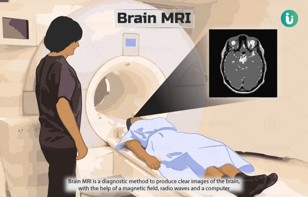
Magnetic resonance imaging (MRI) of the brain is a diagnostic method to produce clear images of the brain, with the help of a magnetic field, radio waves and a computer.

During an MRI scan, a magnetic field is produced around the patient. The scanner then sends radio waves, which alter the alignment of atoms in their body. When the atoms realign, they send out radio signals that are picked up by the scanner and converted into images of the area examined. A contrast dye may be used to improve the quality of the images.

A brain MRI helps to detect problems in the brain. It is the most sensitive imaging test for the brain and can provide detailed images of areas in the brain that are not seen well on an x-ray or CT (computed tomography) scan.

  1. Who cannot have a brain MRI?
  2. Why is a brain MRI done?
  3. How should I prepare for a brain MRI?
  4. How is a brain MRI done?
  5. How will a brain MRI feel like?
  6. What do the results of a brain MRI mean?
  7. What are the risks and benefits of a brain MRI?
  8. What happens after a brain MRI?
  9. Contrast vs Non-contrast brain MRI
  10. What are the other tests that can be done with a brain MRI?

MRI scan is avoided in the following individuals unless evaluated for safety:

  • Pregnant women, especially in the first trimester 
  • People with metal implants in the body, such as:
    • Ear implants 
    • Aneurysm clips
    • Cardiac defibrillators 
    • Pacemakers
    • Metal coils placed in blood vessels
  • People having metal fragments anywhere in their body
myUpchar doctors after many years of research have created myUpchar Ayurveda Urjas Capsule by using 100% original and pure herbs of Ayurveda. This ayurvedic medicine has been recommended by our doctors to lakhs of people for sex problems with good results.
Long Time Capsule
₹719  ₹799  10% OFF
BUY NOW

A brain MRI is also ordered to detect conditions that affect the brain, such as:

Your doctor may order this test to determine the cause of symptoms, such as:

You do not have to skip meals or medications before the test unless instructed by the doctor. Make sure to wear loose clothing without metal ties on the day of the test. Alternatively, you may be given a hospital gown to wear. 

Inform your doctor if you have a fear of closed spaces. He/she may give you a mild sedative before the test. Read more: Claustrophobia symptoms

Tell your healthcare practitioner and the radiologist if you are pregnant or allergic to the contrast material. Do not carry any metal items inside the MRI room, as it can cause burns. These items include:

  • Mobile phones 
  • Electronic watches 
  • Pens
  • Pocket knives 
  • Eyeglasses
  • Jewellery
  • Watches 
  • Credit cards
  • Hairpins

The following procedure is followed for a brain MRI:

  • A technician will position you on the scan table and place a plastic device around your head.
  • If a contrast injection is required, it will be given through a vein in your arm. 
  • The scan table will slide into the tunnel-like opening of the scanner. 
  • The technologist will control the scanner from another room and take images of your head. 
  • He/she can communicate with you through speakers available inside the scanner. 
  • A call button will be provided in case you have any problems during the procedure. 
  • You will be given a headset to block out noises made by the scanner.
  • Make sure to lie very still during the test, as movements can distort or blur the scan images. 

The test usually takes about 45 minutes.

myUpchar doctors after many years of research have created myUpchar Ayurveda Kesh Art Hair Oil by using 100% original and pure herbs of Ayurveda. This Ayurvedic medicine has been recommended by our doctors to more than 1 lakh people for multiple hair problems (hair fall, gray hair, and dandruff) with good results.
Bhringraj Hair Oil
₹599  ₹850  29% OFF
BUY NOW

The procedure itself is painless; however, some individuals may find it uncomfortable to lie still for a long time.

You may hear intermittent clicking, humming and knocking sounds from the scanner. The technician will give you a headset to cancel the noise.

After the test, you may feel some warmth in the scanned area.

If a contrast dye is injected, you may experience a feeling of coldness, headache, metallic taste in the mouth or nausea after the test. Though, these effects usually last only a few moments.

The following conditions can be diagnosed through a brain MRI:

  • Infection of the brain or skull bones (Read more: Brain infection causes)
  • Bleeding in the brain
  • Brain damage due to injury
  • Brain tissue swelling
  • Multiple sclerosis
  • Brain tumours
  • Fluid collection around the brain
  • Structural abnormalities in the brain
  • Stroke

MRI is associated with a few risks, such as:

  • The strong magnetic field can cause malfunctioning of any implanted medical device.
  • In rare cases, the patient develops an allergic reaction to the contrast dye.
  • The contrast dye may be harmful to people with serious kidney problems.

The benefits of an MRI are:

  • A non-invasive procedure with no radiation exposure 
  • Gives detailed images of the brain that are clearer than those obtained from other imaging studies 
  • Detects abnormalities that may be obscured by bone
  • Detects stroke at a very early stage
myUpchar doctors after many years of research have created myUpchar Ayurveda Urjas Energy & Power Capsule by using 100% original and pure herbs of Ayurveda. This Ayurvedic medicine has been recommended by our doctors to lakhs of people for problems like physical and sexual weakness and fatigue, with good results.
Power Capsule For Men
₹719  ₹799  10% OFF
BUY NOW

You can resume your usual diet and activities after the test. However, if a contrast dye is used, you will be monitored for some time for side effects such as itching, swelling, rashes or difficulty breathing. Inform your doctor if you observe any pain or swelling at the site of injection. If a sedative is given, you will be asked to rest until its effects wear off.

A contrast dye may be used to enhance the image quality of the brain MRI. The dye is usually administered to diagnose certain conditions such as tumour and cerebral abscess; however, it is not needed for the diagnosis of conditions such as an aneurysm.

Due to the use of a dye, there is a risk of allergic reaction from contrast MRI while a non-contrast MRI carries no such risk.

After a contrast MRI, the patient has to be monitored for a while for any side effects from the dye. However, you can directly go home after a non-contrast MRI.

A contrast MRI can be risky for those with kidney problems while it is not the case with a non-contrast MRI.

Additional tests ordered will vary with the condition suspected but may include:

  • Lumbar puncture
  • Nerve function study
  • Carotid ultrasound 
  • Cerebral Angiography
  • Echocardiogram 
  • CT scan

Disclaimer: All results must be clinically correlated with the patient’s complaints to make a complete and accurate diagnosis. The above information is provided from a purely educational point of view and is in no way a substitute for medical advice by a qualified doctor.

References

  1. Johns Hopkins Medicine [Internet]. The Johns Hopkins University, The Johns Hopkins Hospital, and Johns Hopkins Health System; Magnetic Resonance Imaging (MRI) of the Spine and Brain
  2. Nemours Children’s Health System [Internet]. Jacksonville (FL): The Nemours Foundation; c2017; Magnetic Resonance Imaging (MRI): Brain
  3. Wilkinson ID, Graves MJ. Magnetic resonance imaging. In: Adam A, Dixon AK, Gillard JH, Schaefer-Prokop CM, eds. Grainger & Allison's Diagnostic Radiology. 6th ed. Philadelphia, PA: Elsevier Churchill Livingstone; 2015:chap 5
  4. Chernecky CC, Berger BJ. Magnetic resonance imaging (MRI) - diagnostic. In: Chernecky CC, Berger BJ, eds. Laboratory Tests and Diagnostic Procedures. 6th ed. St Louis, MO: Elsevier Saunders; 2013:754-757
  5. Aygun N, Zinreich SJ. Overview of diagnostic imaging of the head and neck. In: Flint PW, Haughey BH, Lund V, et al, eds. Cummings Otolaryngology. 6th ed. Philadelphia, PA: Elsevier Saunders; 2015:chap 8.
  6. Petersen RC, Lopez O, Armstrong MJ, Getchius TSD, Ganguli M, Gloss D, Gronseth GS, Marson D, Pringsheim T, Day GS, Sager M, Stevens J, Rae-Grant A. Practice guideline update summary: Mild cognitive impairment: Report of the Guideline Development, Dissemination, and Implementation Subcommittee of the American Academy of Neurology. Neurology. 2018 Jan 16;90(3):126-135. PMID: 29282327
  7. Knopman DS. Cognitive impairment and other dementias. In: Goldman L, Schafer AI, eds. Goldman-Cecil Medicine. 26th ed. Philadelphia, PA: Elsevier; 2020:chap 374
  8. Daroff RB, Jankovic J, Mazziotta JC, Pomeroy SL, eds. Bradley's Neurology in Clinical Practice. 7th ed. Philadelphia, PA: Elsevier; 2016
  9. Skinner S. MRI brain imaging. Aust Fam Physician. 2013 Nov; 42(11): 794-7. PMID: 24217100.
Read on app
cross
Ask your health query from live doctors now!