Skeletal Dysplasia

Dr. Suvansh Raj NirulaMBBS

February 23, 2021

February 23, 2021

Skeletal Dysplasia
Skeletal Dysplasia

Skeletal dysplasias, also called osteochondrodysplasias, is a group of hundreds of disorders with different genetic causes that affect the bone and cartilage growth and development of the skeletal system. While the presentation varies based on the type of skeletal dysplasia, broadly speaking, bone and cartilage growth abnormalities cause distortion of skeleton shape and size. Furthermore, the long bones, spine and skull become disproportionate. The severity of skeletal dysplasia can range from a complete absence of growth, resulting in a fatality, to mild dysfunction, resulting in dwarfism. While these exceptionally rare diseases are marked by significantly short stature, the distinguishing feature of skeletal dysplasia is the disproportionate growth between different body parts. Although primarily a dysfunction of bone and connective tissue growth, other systems like neurological, respiratory and cardiac can be involved as well. Most cases of skeletal dysplasia are caused due to the expression of specific defective genes, transmitted by the carrier parent, in the baby. Besides clinical presentation and genetic testing, radiological imaging studies are of utmost importance for accurate diagnosis, classification, treatment planning and prognosis.

Types of skeletal dysplasia or osteochondrodysplasia

Skeletal dysplasia is classified according to the type of genetic defect or the presenting clinical features. While it is not possible to describe each class of osteochondrodysplasia within this article, naming of the different kinds, depending on the anatomical region affected, is explained below:
  • The prefix ‘acro-’ denotes that the distal parts are affected, i.e. the hands and feet.
  • ‘Meso-’ indicates “middle portion” or ulna and radius bones in the forearm and tibia and fibula in the leg.
  • ‘Rhizo-’ at the beginning of the name implies that the proximal portion of the limb, like the humerus in the upper limb and femur in the lower limb, are implicated.
  • ‘Spondylo-’ in the osteochondrodysplasia term refers to the impacted spinal column.
  • ‘Epi-’ in the name denotes the affection of epiphysis of the bone involved.
  • ‘Meta-’ points to the metaphysis of the affected bone or bones.

Understanding the naming of skeletal dysplasia can help anticipate the signs and symptoms and their management. Furthermore, skeletal dysplasias can be described on the basis of their severity or prognostic outlook as follows:

  • Usually fatal: Achondrogenesis, thanatophoric dysplasia, osteogenesis imperfecta type II
  • Often fatal: Asphyxiating thoracic dystrophy (Jeune's syndrome)
  • Occasionally fatal: Diastrophic dysplasia, metatropic dwarfism, etc.

The four most common types of skeletal dysplasia are:

  • Thanatophoric dysplasia: A severe, usually fatal, type of skeletal dysplasia in which the babies have extremely short limbs with redundant folds of skin, short ribs, narrow chest, enlarged head and forehead, widespread eyes and underdeveloped lungs. Such infants are usually stillborn. Even if they survive into early childhood with intensive medical care, the prognosis is not good.
  • Achondroplasia: Normally, the development of some bones in the body is such that they are present as cartilage in the fetus and morph into bones with advancing maturity. However, in genetically inherited achondroplasia, such change from cartilage to bone, is inhibited and dwarfism ensues in the child. Usually, parents are of average height but the child has a short stature, with a prominently large head and short limbs.
  • Osteogenesis imperfecta: This is also called the brittle bone disease as the bones formed in individuals suffering from this disease are not strong enough and break easily. It arises due to a defect in type 1 collagen, which is a connective tissue protein present in various parts of the body (most significantly, the bones). Other accompanying signs of osteogenesis imperfecta are blue sclera (white portion of the eyeball), loose joints, hearing loss and short height. Cervical artery dissection and aortic dissection can be possible complications. The disease can range from mild to severe.
  • Achondrogenesis: A typically lethal form of skeletal dysplasia in which the infants are either born prematurely or stillborn and present with a small body and other skeletal abnormalities. These babies usually succumb to respiratory failure at, or soon after, birth.
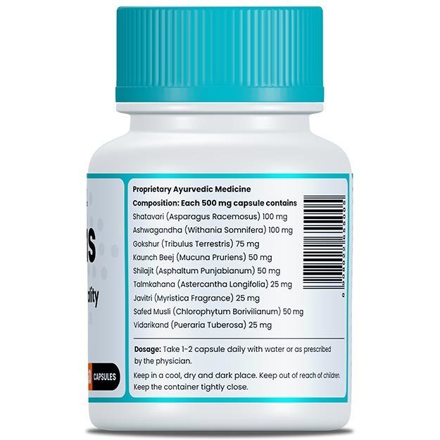
myUpchar doctors after many years of research have created myUpchar Ayurveda Urjas Capsule by using 100% original and pure herbs of Ayurveda. This ayurvedic medicine has been recommended by our doctors to lakhs of people for sex problems with good results.
Long Time Capsule
₹719  ₹799  10% OFF
BUY NOW

Signs and symptoms of skeletal dysplasia

Depending on the type of skeletal dysplasia, the presentation varies. Usually, the bones are misshapen, disproportionate to the rest of the body and the individual has a short stature.

  • Disproportionately short stature (short limbs or short trunk)
  • Disproportionate shortening may be seen in:
    • Short proximal segments (‘Rhizo-’)  - humerus, femur: for example, achondroplasia, hypochondroplasia, diastrophic dysplasia and congenital short femur.
    • Short middle segments (‘Meso-’) - radius, ulna, tibia, fibula: for example, Robinow's syndrome, etc.
    • Short distal segments (‘Acro-’) - metacarpals, phalanges: for example, acrodysostosis and peripheral dysostosis.
    • Short middle and distal segments (‘Acromeso-’) - forearms, hands: for example, acromesomelic dysplasia.
    • Shortening of extremities involving an entire limb: for example, achondrogenesis, etc.
    • Short trunk (short neck, small chest and protruding abdomen): for example, Morquio's syndrome.
  • Some other accompanying features could include:
  • Systemic involvement: Some types of osteochondrodysplasia present with specific signs and symptoms that are:
    • Neurological: Neurological deficits as seen with hydrocephalus or spinal syndromes
    • Cardiac: Aortic dissection, cervical artery dissection, etc.
    • Respiratory: Respiratory failure, etc.

In lethal varieties of osteochondrodysplasia, the child may be stillborn or might succumb to respiratory failure upon birth.

Cause of skeletal dysplasia

Skeletal dysplasia is passed on genetically from parents to their children. This may occur even if the parents do not suffer from it themselves. The usual genetic mutations impact the bone and cartilage growth.

Risk factors of skeletal dysplasia are:

  • Family history of skeletal dysplasia
  • Short stature of parents
  • Maternal hydramnios

Diagnosis of skeletal dysplasia

History: The doctor begins the assessment by carefully taking a thorough medical history. Emphasis is paid to family history, pregnancy and childbirth history as well as the developmental history of the child. While some types of skeletal dysplasia might be diagnosed at birth or soon after, many are suspected later when the child’s stature lags behind that of his or her peers.

Physical examination: A complete physical examination is conducted to detect misshapen body parts, measure the growth (through anthropometry) or even rule out other causes of short stature. Neurological, cardiovascular and respiratory system examination is important.

Anthropometric measurements: Anthropometric refers to the serial measurement of body part circumferences, lengths, weight and height and is initiated in all children at birth. Routinely, the following are recorded:

  • Head circumference
  • Chest circumference
  • Abdominal circumference
  • Weight
  • Body length (up to 2 years of age, thereafter it is replaced by standing height measurements)
  • Height (children older than 2 years of age)
  • Upper segment to lower segment ratio
  • Body mass index (BMI) in older children

These measurements are plotted on standard growth charts to assess whether the baby is growing normally. Any deviation from the expected values for the given age and sex indicate growth abnormalities and may need to be followed up with other studies.

(Read more: Baby development in the first month after birth)

myUpchar doctors after many years of research have created myUpchar Ayurveda Kesh Art Hair Oil by using 100% original and pure herbs of Ayurveda. This Ayurvedic medicine has been recommended by our doctors to more than 1 lakh people for multiple hair problems (hair fall, gray hair, and dandruff) with good results.
Bhringraj Hair Oil
₹599  ₹850  29% OFF
BUY NOW

Differential diagnosis for skeletal dysplasia

Skeletal dysplasia associated dwarfism is typically disproportionate. In case the short stature and shorter bone length are proportionate, the underlying cause may not be skeletal dysplasia but one of the following:

Tests for skeletal dysplasia

Investigations required to diagnose skeletal abnormalities may include:

  • Blood tests: Although skeletal dysplasia is a clinical and radiological diagnosis, many of the other causes of short stature are ruled out by laboratory investigations.
  • Urinalysis: can be helpful in diagnosing urinary tract infections associated with chronic kidney disease
  • Antenatal investigations during pregnancy:
    • Ultrasound: Routine ultrasounds done during pregnancy can detect certain types of skeletal dysplasia in the foetus. If skeletal dysplasia is suspected, the ultrasound is followed up by targeted imaging for fetal anomalies by a more specialised scan. 
    • Genetic counselling and DNA testing: Ultrasonographic anomalies may necessitate the need for other tests, such as amniocentesis, for genetic testing and definitive diagnosis. 
  • Radiological imaging:
    • X-rays: Plain radiographs or X-rays are the first line of imaging used to study and analyse the underlying bone defects. Various specific signs are present in the case of different types of skeletal dysplasia. 
    • CT scan or MRI: It can help further study skeletal abnormalities dented on X-rays. 
    • Brain imaging: Various modalities, X-ray, CT scan or MRI can be used to diagnose tumours producing hormonal imbalance and short stature.

Management of skeletal dysplasia

Management of skeletal dysplasia depends on the type of disease and is usually an amalgamation of medical, orthopaedic, dental and surgical treatment. At times this is followed by physiotherapy, occupational and psychological therapy. Although short stature cannot be improved, the other associated deformities and symptoms can be addressed to a certain degree.

Orthopaedic: Use of braces to prevent bowing of back due to scoliosis or kyphosis.

Dental: Orthodontic treatment with dental braces to correct overcrowding of teeth.

Surgical: Surgical options may include:

  • Correction of cleft palates
  • Correction of deformities like clubfoot
  • Hydrocephalus might need draining of extra fluid from the brain
  • Spinal cord compression due to spinal abnormalities may be relieved
  • Tracheostomy to help respiration can be needed in severe cases

Physiotherapy: Physiotherapy may be recommended with the aim to:

  • Improve muscle strength
  • Increase the range of motion of joints and limbs
  • Promote physical activity and prevent obesity

Psychological: Short stature or disproportionality of body parts can lead to undue mental duress, body image issues and low self-esteem. Psychological therapy can help the individual gain confidence and lead a healthy productive life.

myUpchar doctors after many years of research have created myUpchar Ayurveda Urjas Energy & Power Capsule by using 100% original and pure herbs of Ayurveda. This Ayurvedic medicine has been recommended by our doctors to lakhs of people for problems like physical and sexual weakness and fatigue, with good results.
Power Capsule For Men
₹719  ₹799  10% OFF
BUY NOW

Prevention of skeletal dysplasia

In the case of family history of growth disorders or pre-diagnosed skeletal dysplasia, genetic counselling and testing should be sought by expecting parents. However, sometimes children with skeletal dysplasia may be born to parents of average height without obvious growth disorders.

Prognosis of skeletal dysplasia

The long term outcome of skeletal dysplasia depends on the underlying cause and type of disease. While nearly half of the fetuses with lethal osteochondrodysplasias (like thanatophoric dysplasia) either are stillborn or die prematurely, many children with sub-lethal varieties survive into early childhood. In relatively benign cases, many individuals reach adulthood and lead normal lives.