Download App


What is a Renal Function test (KFT)?
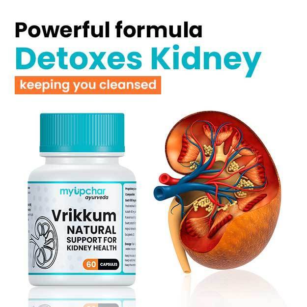
Renal or kidney function tests help to determine whether the kidneys are functioning properly. Kidneys are the main excretory organs of the body, through which metabolic waste, i.e., waste generated by different cells of the body is removed. Specialised kidney cells, called nephrons, can filter waste products, toxic substances and water from blood to form urine, while at the same time reabsorb useful substances selectively.
Creatinine, a by-product of muscle breakdown, and urea, a nitrogen-containing compound are some of the main waste products excreted by kidneys. Blood urea nitrogen (BUN) and serum creatinine test are simple tests that help find out whether nephrons can filter creatinine and urea effectively, thereby, helping assess the extent of kidney damage.
Creatinine clearance is also an important parameter that helps to determine the functioning of kidneys. Its value is derived from the value of serum creatinine.
Furthermore, serum creatinine can be used to determine glomerular filtration rate (GFR). GFR is the amount of filtrate formed in both kidneys each minute and is helpful in diagnosing kidney failure.
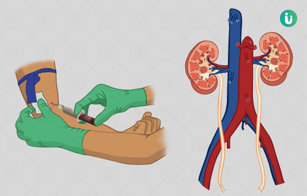
Other than blood tests, imaging tests such as ultrasonography (USG) or computed tomography (CT) scan of kidneys may also be advised to obtain a clear image of the shape and size of kidneys. It helps determine the presence of stones, tumours or obstructions in any part of kidneys. Testing of urine may also be performed to gain an understanding of the excretory ability of kidneys in certain disease conditions.
- Why is Renal Function test (KFT) performed?
- How do you prepare for a Renal Function test (KFT)?
- How is Renal Function test (KFT) performed?
- Renal Function test (KFT) results and normal values/range

Why is Renal Function test (KFT) performed?
Blood tests such as BUN and serum creatinine and urine tests may be performed if the individual shows symptoms such as lower back pain; swelling of ankles, wrists, face or abdomen; decreased frequency and quantity of urine through the day; burning sensation during urination; dehydration; fatigue; loss of appetite; nausea; vomiting; or persistent high blood pressure or fall in blood pressure while changing positions.
Imaging tests may be performed to evaluate symptoms of kidney stones such as intense sharp pain in the abdomen other than the above-mentioned classic symptoms of kidney problems or to assist in the placement of a needle while taking a kidney tissue for biopsy.


How do you prepare for a Renal Function test (KFT)?
No special preparation or precaution is required for these tests.
The physician must be informed regarding the consumption of drugs such as cimetidine or antibiotics such as trimethoprim that interfere with the test results.
How is Renal Function test (KFT) performed?
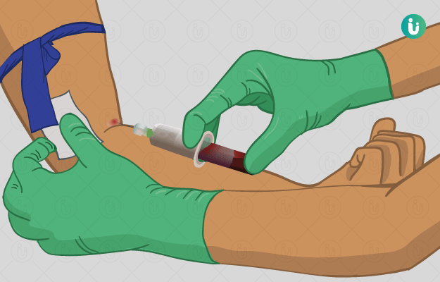
- Blood test: A tourniquet may be tied onto your arm, and a small amount of blood will be withdrawn from a vein with a sterile needle after swabbing the concerned area with an appropriate antiseptic. The sample is then collected into a container. Some individuals may experience light-headedness after blood collection.
- Urine test: A specimen cup will be provided to the individual, and he or she will be asked to collect their urine in it. The specimen must be sent to the laboratory within one hour of collection.
- Imaging tests:
- Ultrasonography (USG): High-frequency sound waves are used to generate the image of kidneys onto a monitor screen.
- CT scan: A CT scan machine takes x-ray images of kidneys from different angles to determine any abnormalities in organs.
Renal Function test (KFT) results and normal values/range
Normal Results
Blood tests:
- Serum creatinine:
- Men: 0.7 to 1.3 mg/dL (61.9 to 114.9 µmol/L)
- Women: 0.6 to 1.1 mg/dL (53 to 97.2 µmol/L)
- BUN: 6 to 20 mg/dL
- Creatinine clearance: 120-140 mL/min
- GFR: 120 mL/min
Urine test:
- Appearance: Normal urine will appear pale yellow and should be clear.
- Chemical: Results should be negative for ketones, leucocyte esterase, bilirubin, urine bilirubin and nitrites. Glucose in urine should not be over 130 mg/dL, and protein should not be over 150 mg/dL.
- Microscopic: The urine sample should be free from crystals, bacteria and yeast. Red blood cell (RBC) and white blood cell (WBC) content must be less than 2 RBC’s or 2-5 WBC's/HPF, respectively.
- Imaging tests: USG or CT scan of kidneys gives a detailed description of the size and shape of each anatomical part of the urinary system such as the right and left kidneys, ureters, urinary bladder and urethra.
Abnormal Results
Blood tests:
- Serum creatinine:
Higher-Than-Normal serum creatinine may be due to a blockage in the urinary tract, acute or chronic kidney failure, decreased blood flow to kidneys, dehydration, higher-than-normal breakdown of muscle fibres known as rhabdomyolysis, or even due to an adverse reaction to certain antibiotics and drugs.
Lower-than-normal serum creatinine may be due to diseases and conditions that lead to decreased muscle mass such as AIDS, cancer, congestive heart failure, osteoporosis, malnutrition and many others.
- BUN:
Higher-than-normal BUN reflects kidney diseases such as acute tubular necrosis, glomerulonephritis and pyelonephritis, congestive heart failure, gastrointestinal bleeding, chronic kidney failure(which may or may not require dialysis) dehydration or urinary tract obstruction.
Lower-than-normal BUN reflects malnutrition, low-protein diet, liver disease and too much hydration.
- Creatinine clearance:
Lower-than-normal creatinine clearance may indicate damage to nephrons, decreased blood flow to kidneys, heart failure, kidney failure, dehydration or obstruction at the outlet of the bladder.
- GFR:
A GFR of 90 mL/min or higher indicates normal kidney function or stage 1 chronic kidney disease (CKD).
GFR of 60-89 mL/min indicates stage 2 CKD with mild loss of kidney function.
Stage 3A CKD will have a mild-to-moderate loss of kidney function with GFR of 45-59 mL/min.
Stage 3B CKD will have a moderate-to-severe loss of kidney function with GFR of 30-44 mL/min.
Stage 4 CKD will have severe loss of kidney function with GFR of 15-30 mL/min and a GFR of lower than 15 mL/min indicates complete kidney failure.
Urine test:
- Appearance: Cloudy urine implies an underlying infection. Red or brown urine indicates a UTI or acute kidney injury. Sometimes urine may also appear orange due to anti-tuberculosis drugs like rifampicin.
- Chemical:
- A positive ketone test implies a serious condition called diabetic ketoacidosis. Further metabolic tests such as serum sodium, potassium, calcium and chloride will be ordered. (Read more: Electrolytes test)
- Presence of leucocyte esterase and nitrites in the urine indicate a bacterial infection.
- High level of glucose in urine is seen in diabetes mellitus and is also common in pregnancy.
- Higher-than-normal level of protein in urine is seen in diabetes mellitus, fever, nephritic syndrome, multiple myeloma and many other conditions.
- Microscopic:
- Presence of RBCs could indicate conditions like sickle cell anaemia, renal cell carcinoma, benign prostatic hyperplasia, kidney stones and others.
- Presence of excess amount of WBCs could indicate that the body is fighting an infection, and the presence of crystals could indicate gout or kidney stones.
- Imaging tests: These tests will give the physician a clear idea about structural abnormalities, tumours, obstructions, stones and any improper blood supply to the kidney.
Disclaimer: All results must be clinically correlated with the patient’s complaints to make a complete and accurate diagnosis. The above information is provided from a purely educational point of view and is in no way a substitute for medical advice by a qualified doctor.

Doctors for Kidney / Renal Function Test

Dr. Anvesh Parmar
Nephrology
12 Years of Experience
DR. SUDHA C P
Nephrology
36 Years of Experience

Dr. Mohammed A Rafey
Nephrology
25 Years of Experience

Dr. Soundararajan Periyasamy
Nephrology
30 Years of Experience
References
- © 2019 National Kidney Foundation [internet]; Tests to Measure Kidney Function, Damage and Detect Abnormalities
- Roger Walker, Cate Whittlesea. Clinical Pharmacy and Therapeutics. Acute kidney injury. 5th Edition. 2012. 255-269.
- Tomasz Szopiński,corresponding, Elżbieta Keller, and František Záťura. Kidney ultrasound – what is important for a urologist?. J Ultrasonv.16(67); 2016 Dec PMC5269524
- MedlinePlus Medical Encyclopedia: US National Library of Medicine; Creatinine blood test
- MedlinePlus Medical Encyclopedia: US National Library of Medicine; Creatinine clearance test
- Chernecky CC, Berger BJ. Creatinine clearance - serum and urine. In: Chernecky CC, Berger BJ, eds. Laboratory Tests and Diagnostic Procedures. 6th ed. St Louis, MO: Elsevier Saunders; 2013:401-403
- Inker LA, Fan L, Levey AS. Assessment of renal function. In: Johnson RJ, Feehally J, Floege J, eds. Comprehensive Clinical Nephrology. 5th ed. Philadelphia, PA: Elsevier Saunders; 2015:chap 3.
- Oxford handbook of clinical and laboratory investigation. Chapter 13; pg no 809-810.
- Simerville JA, Maxted WC, Pahira JJ. Urinalysis: a comprehensive review.. Am Fam Physician. 2005 Mar 15;71(6):1153-62. PMID: 15791892
- Gerard J. Tortora, Bryan Derrickson. Principles of anatomy and physiology. 14th ed. Wiley Publication; 2014. Chapter 26.7. pg 1008-100
- @ 2019 National Kidney Foundation [internet]; Estimated Glomerular Filtration Rate (eGFR)
- Kalyani RR, Corriere M, Ferrucci L. Age-related and disease-related muscle loss: the effect of diabetes, obesity, and other diseases.. Lancet Diabetes Endocrinol. 2014 Oct;2(10):819-29. doi: 10.1016/S2213-8587(14)70034-8. Epub 2014 Mar 6.
- Gerard J. Tortora, Bryan Derrickson. Principles of anatomy and physiology. 14th ed. Wiley Publication; 2014. Chapter 26;p.994, 1010.
- MedlinePlus Medical Encyclopedia: US National Library of Medicine; BUN - blood test
- National Health Service [internet]. UK; How should I collect and store a urine sample?
- Landry DW, Bazari H. Approach to the patient with renal disease. In: Goldman L, Schafer AI, eds. Goldman-Cecil Medicine. 25th ed. Philadelphia, PA: Elsevier Saunders; 2016:chap 114.