Download App
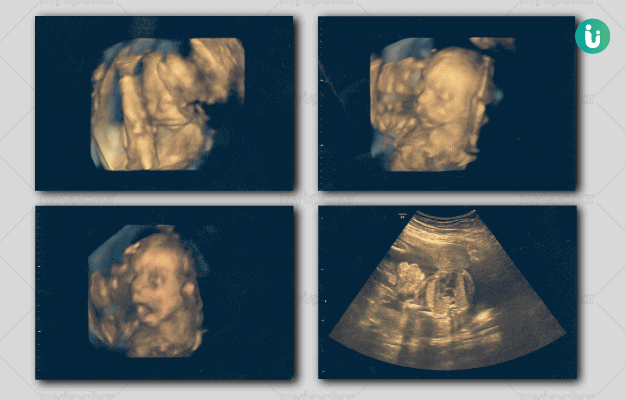


What is 3D Ultrasound?
An ultrasound scan uses high-frequency sound waves to create a picture of internal organs, including the baby and placenta and is a commonly used diagnostic procedure during pregnancy. It is recommended to evaluate foetal growth and development and also to find the due date of delivery. An ultrasound helps the parents bond with the unborn baby by allowing them to view images of the baby and listening to its heartbeat.
Different types of ultrasound procedures exist with the same basic procedure. An ultrasound 3D scan utilises specially designed transducer probes along with a special software to create 3D pictures of the baby in the womb. Unlike the normal or 2D ultrasound images, this scan provides some width, height and depth to the image, helping view some facial features of the baby and parts of the body like fingers and toes in much more detail.
- Why is 3D Ultrasound scan done during pregnancy?
- How do you prepare for 3D ultrasound scan?
- How is 3D ultrasound scan performed during pregnancy?
- What do the results of 3D ultrasound scan during pregnancy mean?
- How much does a 3D Ultrasound scan during pregnancy costs?

Why is 3D Ultrasound scan done during pregnancy?
A 3D ultrasound is recommended when more than just a flat 2D picture is needed. It helps in the diagnosis of diseases and abnormal conditions of the foetus.
An ultrasound scan is ordered at different times during the pregnancy for various purposes. This test is recommended in the first trimester:
- For the diagnosis of ectopic pregnancy, a complication in pregnancy in which the embryo attaches outside the uterus
- To identify placental structure
- To establish the important dates in pregnancy, e.g., age of the foetus at the time of the test
- To examine the structures in the pelvis
- To determine the number of foetuses
In the second trimester, this test is recommended:
- To monitor the growth of the baby
- To identify the presence of abnormalities in foetus
- To evaluate blood flow patterns
- To observe the behaviour and activity of the foetus
- To determine the amount of amniotic fluid in the womb
- To examine the placenta
- To measure the length of the cervix
In the third trimester, this test is recommended:
- To monitor the growth and development of the baby
- To identify baby’s position in the womb
- To assess the placenta
- To determine the amount of amniotic fluid in the womb

How do you prepare for 3D ultrasound scan?
An ultrasound-pregnancy 3D scan does not require any special preparation. However, the doctor may sometimes recommend drinking about 4 to 6 glasses of water so that the bladder is a full and clearer view of the baby can be obtained. Also, wearing a hospital gown is advised for this procedure.
How is 3D ultrasound scan performed during pregnancy?
During pregnancy, ultrasound 3D scan procedure can be done in two ways:
- Transvaginal ultrasound: In this test, a small transducer probe is inserted into the vagina, allowed to rest against vaginal wall, and images are captured. It produces sharper images and is generally ordered during the early phases of pregnancy.
- Abdominal ultrasound: In this test, a gel is applied on the abdomen, and the ultrasound transducer probe is glided over the belly to produce an image. This procedure needs a full bladder for better imaging; therefore, water consumption is needed before this test.
The test is non-invasive and does not pose any risk to the mother or the baby. However, the probe puts some pressure in the vagina or on abdominal region, which may cause slight discomfort to the mother.
Although the test is said to be free of any risks, long-term effects of prolonged or repeated exposure to ultrasound are unclear. It is advised to get this test done only when recommended by the doctor.
What do the results of 3D ultrasound scan during pregnancy mean?
Normal results:
Normal results indicate that the baby is healthy and growing accordingly as per the gestational age. The surrounding structures, placenta and amniotic fluid also appear to be normal. In this case, the doctor advises continuing with usual antenatal care. It is important to note that normal results may vary slightly sometimes; thus, it is best to consult a doctor for correct interpretation of results.
Abnormal results:
Abnormal results may be due to the presence of one of the following conditions:
- Multiple pregnancies
- Miscarriage
- Presence of abnormally high or low levels of amniotic fluid
- Improper position of foetus in the womb
- Slow or poor growth or development of baby
- Ectopic pregnancy
- Birth defects
- Tumours in pregnancy
- Problems of the uterus, ovaries or any other part of the pelvis
Disclaimer: All results must be clinically correlated with the patient’s complaints to make a complete and accurate diagnosis. The above information is provided from a purely educational point of view and is in no way a substitute for medical advice by a qualified doctor.

How much does a 3D Ultrasound scan during pregnancy costs?
The cost of this test ranges from 1,000 to 3,000 INR depending on the laboratory and city it is conducted in.

References
- Stanford Children's Health: Lucile Packard Children's Hospital, Stanford; Ultrasound in Pregnancy
- American Pregnancy Association: Ultrasound: Sonogram
- US Food and Drug Administration (FDA) [internet]; Ultrasound Imaging
- National Health Service [internet]. UK; Ultrasound scans in pregnancy - Your pregnancy and baby guide
- Lazarus E, Levine D. The first trimester. In: Rumack CM, Levine D, eds. Diagnostic Ultrasound. 5th ed. Philadelphia, PA: Elsevier; 2018:chap 30.
- Wapner RJ. Prenatal diagnosis of congenital disorders. In: Creasy RK, Resnik R, Iams JD, Lockwood CJ, Moore TR, Greene MF, eds. Creasy and Resnik's Maternal-Fetal Medicine: Principles and Practice. 7th ed. Philadelphia, PA: Elsevier Saunders; 2014:chap 30.
- Richards DS. Obstetric ultrasound: imaging, dating, growth, and anomaly. In: Gabbe SG, Niebyl JR, Simpson JL, et al, eds. Obstetrics: Normal and Problem Pregnancies. 7th ed. Philadelphia, PA: Elsevier; 2017:chap 9.
- Wolf RB. Abdominal imaging. In: Creasy RK, Resnik R, Iams JD, Lockwood CJ, Moore TR, Greene MF, eds. Creasy and Resnik's Maternal-Fetal Medicine: Principles and Practice. 7th ed. Philadelphia, PA: Elsevier Saunders; 2014:chap 24.