Download App

What is Chandler’s syndrome?
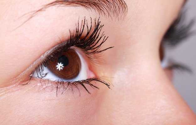
Chandler’s syndrome is a condition of the eye where there is swelling of the cornea, disfigurement of the iris and unusually high pressure within the eye. It belongs to a triad of eye disorders called iridocorneal syndrome where the corneal endothelium (a thin tissue of the cornea) becomes abnormal and appears like ‘hammered silver’. It affects females more than males and is common in young to middle-aged adults.
What are its main signs and symptoms?
The main signs and symptoms include:
- Blurred vision
- Swelling of the cornea
- Abnormal iris
- Rainbow-coloured halos around lights
- Tunnel vision
The pupil of the eye appears displaced from its normal position and distorted in shape and size. The iris size reduction is mild compared to other similar eye disorders. The abnormal corneal endothelium is seen as a hammered silver surface behind the cornea.
Similar symptoms may be seen in the other two variants of the iridocorneal syndrome, namely:
- Progressive iris atrophy
- Cogan-Reese syndrome
Glaucoma or increased pressure within the eye has a prevalence as high as 82% in patients with Chandler’s syndrome.
What are its main causes?
The exact cause is yet unknown. Some suspect inflammation or chronic viral infection as the probable cause. The endothelial surface usually pumps the aqueous humour from the cornea. When this action fails, fluid accumulation in the cornea occurs and results in blurry vision. This can lead to glaucoma, which presents as headache and blurring of vision.
How is it diagnosed and treated?
The condition typically requires careful evaluation and a full ophthalmic examinationto be diagnosed. Chandler’s syndrome is usually diagnosed in patients with a condition called as unilateral, secondary angle-closure glaucoma. Differential diagnosis is done to rule out other disorders with a similar presentation. Specific tests include:
- Gonioscopy
- Intraocular pressure and corneal thickness measurement
- Visual field test
- Optic nerve imaging
Glaucoma evaluation is usually advised to assess the swelling and pressure in the eye.
The treatment lies in reduction of the swelling which increases the pressure in the eye. Topical medicines are used as first-line treatment. Eyedrops to reduce pressure in the eyes may be prescribed. Milder cases can be managed with the use of soft contact lenses and hypertonic saline solutions.
Surgical methods include:
- Trabeculectomy to reduce pressure in the eye
- Corneal transplant
Outcomes of the disorder depends on the development of complications, which, in turn, depends on the timing of diagnosis and success or failure of treatment. A glaucoma specialist can assess the condition and formulate a treatment plan to improve vision and provide better outcomes.
Self-care tips include:
- Avoid strain to the eyes.
- Yoga, meditation and recreational activities can ease the strain to the eyes.
- Regular exercise can lower eye pressure and minimise chances of loss of vision.
- Dietary supplements essential for the eyes include vitamins A, vitamin E and vitamin C, zinc, copper and selenium.
- Regular eye check-ups need to be done to prevent further disease progression.