What is a Four-Dimensional Scan?

Four-dimensional (4D) ultrasound is an imaging technique. It is used worldwide in the field of obstetrics and gynaecology for prenatal imaging of the foetus in mother’s womb. This scan helps in real-time visualisation of the movements of a baby in the womb. It is different from 2D and 3D ultrasound scans, as it gives a complete assessment of the foetus during pregnancy with high-definition clarity. The technique makes use of high-frequency sound waves that bounce off the internal organs and generates images. 

4D scan also captures foetal movements and is also effective in the early diagnosis of prenatal neurodevelopment problems in foetus.

This ultrasound scan is sometimes used along with computerised tomography (CT) in case of certain types of cancers, especially to examine the movement of tumours in lungs with respect to the respiration of the patient.

  1. Why is a 4D Scan performed?
  2. How do you prepare for a 4D Scan?
  3. How is a 4D Scan performed?
  4. What do 4D Scan results indicate?

A 4D ultrasound scan has become a universal choice among gynaecologists all over the world in diagnosing prenatal problems. It enables gynaecologists to examine the foetus much more clearly than a normal 2D or 3D scan.

A 4D scan can be advised in all trimesters of pregnancy, particularly in the second and third, for the following assessments: 

  • General movements during the first trimester, which include upper limb movements, head rotation and stretching movements
  • During the second and third trimesters for facial expressions, such as eye blinking, yawning, sucking and swallowing
  • Recording of complex movements, such as hand to eye, hand to mouth and hand to head in detail

The images obtained help in accessing the development of the central nervous system of the foetus; those with detected anomalies using the scan should be screened further.

Assessments of the images obtained using 4D ultrasound are subjected to the Kurjak’s antenatal neurodevelopmental test (KANET) by many gynaecologists. KANET is a scoring system designed to determine the abnormalities in foetus during the third trimester. It evaluates the qualitative and quantitative aspects of foetal neurobehavioral development. Foetal observable movement system (FOMS), may further provide an insight into the neurological behaviour of the foetus.

(Read more: Foetal development month by month during pregnancy)

myUpchar doctors after many years of research have created myUpchar Ayurveda Urjas Capsule by using 100% original and pure herbs of Ayurveda. This ayurvedic medicine has been recommended by our doctors to lakhs of people for sex problems with good results.
Long Time Capsule
₹719  ₹799  10% OFF
BUY NOW

A 4D scan using ultrasound is a painless and non-invasive procedure and requires no special preparations. The patient is asked to drink 4-6 glasses of water prior to the test for a better view of the foetus and is allowed to empty their bladder only after the test is completed. Fasting is not required before this test.

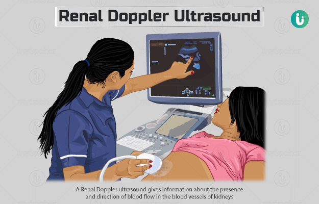
The patient is made to lie down on a table, and a gel is applied to their abdomen so the transducer can glide over the skin softly. When the transducer touches the skin, it sends high-frequency sound waves, and the images of the internal organ are visible on a computer screen. The gel is wiped off after the test. A skilled radiologist makes a note of the findings from the images.

The images can be printed and made available to the gynaecologist. 4D scan lasts for almost 30 minutes, and the results are available on the same day. The only discomfort one may experience is due to fullness of the bladder.

myUpchar doctors after many years of research have created myUpchar Ayurveda Kesh Art Hair Oil by using 100% original and pure herbs of Ayurveda. This Ayurvedic medicine has been recommended by our doctors to more than 1 lakh people for multiple hair problems (hair fall, gray hair, and dandruff) with good results.
Bhringraj Hair Oil
₹599  ₹850  29% OFF
BUY NOW

Normal results: Interpretation of 4D ultrasound scan results is based on the qualitative monitoring of foetal movements and behaviour. However, some gynaecologists may also use the KANET score, which is based on both qualitative and quantitative assessments.

Normal results indicate proper movements of the head, trunk and neck, which appear smooth and show an increase and decrease in the amplitude.

These movements last for 20 seconds. A KANET score of 14-20 is normal.

Abnormal results: If foetal movements are repetitive, monotonous and chaotic and involve contractions of muscles, the results are considered abnormal.

On a quantitative level, abnormal KANET scores range as 0-5, whereas 5-13 are borderline.

Borderline or abnormal prenatal results are indicative of the need for a post-natal test, and the scores are compared. Infants born thereafter should be closely observed for 2 years, as they may suffer from neurological disorders.

Sometimes, a gynaecologist may advise termination of pregnancy due to abnormal results. Abnormal values suggest the following structural deformities and neurological abnormalities:

  • Cerebral palsy, which is a group of movement disorders
  • Dandy-Walker malformation in which malformation of the cranial, vascular and middle ear occurs
  • Skeletal dysplasia, in which, there are abnormalities of bones and spine
  • Spina bifida, a condition in which spinal cord is not properly formed
  • Encephalocele, where the neural tube fails to close, leading to structural deformities

In a nutshell, a 4D scan uses ultrasound technology for capturing the growth and development of motor and neurological activities of the foetus. It predicts post-natal abnormalities and structural deformities. Repeated 4D scans are necessary to monitor pre-existing foetal conditions.

Disclaimer: All results must be clinically correlated with the patient’s complaints to make a complete and accurate diagnosis. The above information is provided from a purely educational perspective and is in no way a substitute for medical advice from a qualified doctor.  

References

  1. Lebit FD and Vladareanu R. The Role of 4D Ultrasound in the Assessment of Fetal Behaviour. Obstetrics and Gynecology. Maedica A Journal of Clinical Medicine. 2011;6(2):120-130.
  2. Bonilla-Musoles F, Bonilla Jr F, Raga F, Caballero O, Cadete C, and Machado LE. Second trimester anomaly scan using 3D/4D. Donald School Journal of Ultrasound in Obstetrics and Gynecology. 2015;9(4):372-381.
  3. Slotman BJ, Lagerwaard FJ, and Senan S. 4D imaging for target definition in stereotactic radiotherapy for lung cancer. Acta Oncologica. 2006;45:966-972.
  4. Kurjak A, Predojević M, and Kadić AS. Fetal Behavior in 4D ultrasound in the progress of perinatal medicine. J Health Med Informat S11: 006. doi:10.4172/2157-7420.S11-006 P
  5. Kurjak A. The normal and abnormal fetal neurodevelopment assessed by new test – kanet. DOI: 10.5644/PI2015-159.02 Accessed on 29 May 2019.
  6. Toshiyuki HJ. Current status of fetal neurodevelopmental assessment: Four dimensional ultrasound study. Obstet. Gynaecol Res. 2016;42(10):1211–1221.
Read on app
cross
Ask your health query from live doctors now!