What is a Renal Doppler Ultrasound?

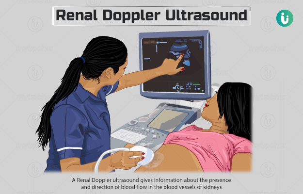
A Renal Doppler ultrasound gives information about the presence and direction of blood flow in the blood vessels of kidneys (renal blood vessels).

Doppler ultrasound is a type of ultrasound that uses sound waves to create an image of the blood flow in your major blood vessels, something that you can’t see on a regular ultrasound.

There are different types of Doppler ultrasound tests, namely, colour Doppler, power Doppler, spectral Doppler, duplex Doppler and continuous wave Doppler.

  1. Who cannot have a Renal Doppler Ultrasound?
  2. Why is a Renal Doppler Ultrasound done?
  3. How should I prepare for a Renal Doppler Ultrasound?
  4. What is the procedure for a Renal Doppler Ultrasound?
  5. How will a Renal Doppler Ultrasound feel?
  6. What do the results of a Renal Doppler Ultrasound mean?
  7. What are the risks and benefits of Renal Doppler Ultrasound?
  8. What happens after the Renal Doppler Ultrasound?
  9. What are the other tests that can be done with a Renal Doppler Ultrasound?

There are no contraindications for this test. This test is also ordered during pregnancy.

Healthcare practitioners may order this test for the following purposes:

  • To look for renal hypertension, especially when the results of an angiogram test are ambiguous. An angiogram is an x-ray test that determines blockages in blood vessels). Renal hypertension refers to high blood pressure that occurs due to stenosis (narrowing) in arteries of kidneys. 
  • As a follow-up to angioplasty, a procedure to open blocked blood vessels.
  • For the diagnosis of renal vein thrombosis (formation of a clot in a blood vessel that carries blood away from the kidney) in infants. This may develop as a complication of dehydration, and the baby would show symptoms like reduced frequency of urination and discoloured/cool hands and feet.
  • For diagnosis of renal vein thrombosis in individuals with the following conditions:
    • Kidney tumour: A blood clot may occur due to a tumour in the kidney. Patients with tumour show symptoms such as loss of appetite, blood in urine, weight loss and anaemia.

      If you are tired of dieting and exercising and are not able to lose weight, then use myUpchar Ayurveda Medarodh Fat Burner Capsule, it has no side effects, order it today and avail the benefits.
       
    • Hypovolemia: This condition is associated with low blood volume. Hypovolemia symptoms include general weakness, pale skin, sweating and unconsciousness.
  • To evaluate whether the blood flow in kidneys is healthy if a person is suspected to have had trauma to their kidneys.
  • As an alternative to x-ray with contrast dye in patients who for some reason cannot take a contrast dye, to detect obstruction in kidneys. Individuals who are allergic to contrast agents would show symptoms like itchy eyes and hives.

You should not eat/drink anything for a minimum of 8 hours before the test. Nicotine may narrow blood vessels and give false results. Therefore, you may need to stop using nicotine-containing products, like cigarettes and chewing tobacco, two hours before the test.

A Renal Doppler Ultrasound includes the following steps:

  • You may be asked to change into a gown.
  • The radiography staff will ask you to lie on your back on an examination table and will cover you with a blanket except for your abdomen.
  • They will then apply a gel on the front and sides of your abdomen and will hold and move a plastic device called a transducer on top of the gel to obtain the necessary images.
  • Try to remain very still during the procedure. You may hear sounds during the examination which are normal and denote the blood flowing through your blood vessels.
  • You may be asked to hold your breath or roll onto your side during the procedure if required.
  • After the scanning is completed, the staff will provide you with something to wipe off the gel.
  • The procedure may take up to 60 minutes.

Usually, no discomfort is felt during the test. The gel may feel cold unless it is first warmed to body temperature. You will occasionally feel the pressure of the transducer, which can be a little uncomfortable but should not hurt.

The test can be used in the investigation of any of the following conditions:

  • Diagnose the following conditions:
    • Renal artery stenosis and renal hypertension
    • Renal vein thrombosis
    • Obstruction in the kidneys
    • Arteriovenous malformation/fistula: A condition that may result in enlargement of the main blood vessels of the kidney
    • Aortic dissection: Tear in the major blood vessel (aorta) that carries blood out of the heart
    • Aortic aneurysm: A bulge in the aorta
  • Trauma to the kidneys
  • To differentiate between a tumour of the kidneys and pseudotumour (false tumour)

The procedure is painless and non-invasive, with no radiation involved. No risk is associated with this test.

You can leave soon after the test is completed and resume your normal activities.

In some cases, your medical practitioner may order additional tests, such as arteriography, to further evaluate abnormal findings and determine the course of treatment.

Disclaimer: All results must be clinically correlated with the patient’s complaints to make a complete and accurate diagnosis. The above information is provided from a purely educational point of view and is in no way a substitute for medical advice by a qualified doctor.

References

  1. Gameraddin M. Ultrasound of the Kidneys: Application of Doppler and Elastography. In: Gamie Samia Ali Abdo and Foda Enas Mahmoud. Essentials of Abdominal Ultrasound. 2019.
  2. Diagnostic Imaging Pathways [Internet]. Government of Western Australia; Information for Consumers - Ultrasound (Doppler)
  3. National Heart, Lung, and Blood Institute [Internet]. Bethesda (MD): U.S. Department of Health and Human Services; Echocardiography
  4. UVA Health System [Internet]. University of Virginia. US; Ultrasound and Doppler ultrasound
  5. Radiological Society of North America (RSNA) [internet]. Oak Brook. Illinois. USA; General Ultrasound
  6. Stanford Health Care [internet]. Stanford Medicine. Stanford Medical Center. Stanford University. US; Doppler Ultrasound
  7. UF Health [Internet]. University of Florida Health. Florida. US; Duplex ultrasound
  8. UW Health: American Family Children's Hospital [Internet]. Madison (WI): University of Wisconsin Hospitals and Clinics Authority; Doppler Ultrasound
  9. Johns Hopkins Medicine [Internet]. The Johns Hopkins University, The Johns Hopkins Hospital, and Johns Hopkins Health System; Pelvic Ultrasound
  10. Merck Manual Consumer Version [Internet]. Kenilworth (NJ): Merck & Co. Inc.; c2018. Ultrasonography
  11. National Heart, Lung, and Blood Institute [Internet]. Bethesda (MD): U.S. Department of Health and Human Services; Heart failure
  12. The Ohio State University: Wexner Medical Center [Internet]. Ohio. US; Doppler Ultrasound
  13. Mycourses. Harvard Medical School [internet]: Harvard University. US; Menu of Radiologic Tests: Ultrasound (US)
  14. Allan PL, Baxter Grant, Weston Michael. Clinical Doppler Ultrasound. 3rd ed. Philadelphia: Churchill Livingstone Elsevier; 2011.
  15. Pagana K.D, Pagana T.J, Pagana T.N. Mosby’s Diagnostic and Laboratory Test Reference. 14th ed. St louis: Mosby Inc.; 2018.
  16. Urology Care Foundation. American Urological Association [internet]. Maryland. U.S.; What is Kidney (renal) Trauma?
  17. Stanford Health Care [internet]. Stanford Medicine. Stanford Medical Center. Stanford University. US; Renal vein thrombosis
  18. American Academy of Pediatrics [internet]. Illinois. US; Signs of Dehydration in Infants & Children
  19. Groeneveld ABJ. Hypovolemic shock. In: Parrillo JE, Dellinger RP, eds. Critical Care Medicine: Principles of Diagnosis and Management in the Adult. 4th ed. Philadelphia, PA: Elsevier Saunders; 2014:chap 26.
  20. Rivers E. Approach to the patient with shock. In: Goldman L, Schafer AI, eds. Goldman-Cecil Medicine. 25th ed. Philadelphia, PA: Elsevier Saunders; 2016:chap 106.
  21. Puskarich MA, Jones AE. Shock. In: Walls RM, Hockberger RS, Gausche-Hill M, eds. Rosen's Emergency Medicine: Concepts and Clinical Practice. 9th ed. Philadelphia, PA: Elsevier; 2018:chap 6.
  22. den Uil CA, Klijn E, Lagrand WK, et al. The microcirculation in health and critical disease. Prog Cardiovasc Dis. 2008;51(2):161–170. PMID: 18774014.
  23. Tarrant AM, Ryan MF, Hamilton PA, Benjaminov O. A pictorial review of hypovolaemic shock in adults. Br J Radiol. 2008;81(963):252–257. PMID: 18180262.
  24. Society for Vascular Surgery [internet]. US; Angiogram
  25. National Institute of Diabetes and Digestive and Kidney Diseases [internet]: US Department of Health and Human Services; Renal Artery Stenosis
  26. Johns Hopkins Medicine [Internet]. The Johns Hopkins University, The Johns Hopkins Hospital, and Johns Hopkins Health System; Angioplasty and Stent Placement for the Heart
  27. Daviess Community Hospital [internet]. Washington. Indiana. US; Renal Artery/Kidney Doppler Ultrasound
  28. CardioSmart: American College of Cardiology [Internet]. Washinton D.C. US; Doppler Ultrasound
  29. Lee Min Hee, Lee Hae Kyung, Choi Seo-Youn, Yi Boem Ha. The Effectiveness of Real-Time Feedback with an Audible Pulse: A Preliminary Study in Renal Doppler Ultrasonography. PLoS ONE. 2016 Sep; 11(9): e0163953.
  30. HealthyWA [internet]. Department of Health: Government of Western Australia; Doppler ultrasound
  31. Centers for Disease Control and Prevention [internet]. Atlanta (GA): US Department of Health and Human Services; Aortic Aneurysm
Read on app
cross
Ask your health query from live doctors now!