Arteriovenous Malformation

Dr. Nabi Darya Vali (AIIMS)MBBS

April 24, 2020

June 04, 2022

Arteriovenous Malformation

Each part of our body has been given a specific task. For instance, the lungs are for breathing, the heart is for pumping blood and the brain is for managing all the systems of the body.

Similarly, the arteries take oxygen-rich blood from the heart to the rest of the body’s tissues and cells, while the veins return oxygen-depleted blood to the lungs and heart. These arteries and veins are connected to each other with the help of tiny blood vessels called capillaries. 

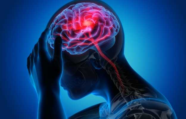
In the absence of adequate capillaries, the arteries and veins can get entangled and form an arteriovenous malformation (AVM). Essentially, in the absence of capillaries, multiple arteries get jumbled up. The blood from these arteries flows directly into a vein, causing its enlargement in some places. This can weaken the blood vessels in the brain.

AVMs are more common in males than in females. A person suffering from an AVM may present with symptoms such as headaches, dizziness, slurred speech, blurred vision, loss of balance and coordination, seizures and stroke.

Doctors do not know the exact cause of AVMs but they are assumed to be a result of genetic mutation. They can be present in a newborn, but they can also develop later in life.

AVMs can be diagnosed with the help of a CT scan, MRI scan, MRA or CT angiogram.

The treatment of AVM includes embolization which helps in reducing the size of the AVM. Followed by a surgery, craniotomy, which is done to remove the AVM from the brain. After the surgery, radiation therapy is given to make sure that the AVM has cleared completely from the brain.

AVMs can be managed easily if diagnosed in the early stages. Here in this article, we will tell you about the symptoms, complications and treatment of arteriovenous malformations or AVMs.

Stages of arteriovenous malformation

Arteriovenous malformations (AVMs) might grow and change over time. According to the Schöbinger staging system, there are four different stages of AVMs; however, not all AVMs go through every stage:

  • Stage I: This stage is also known as quiescence, as the AVM is dormant. At this stage, the skin on top of the AVM feels warm and may appear pink or red in colour.
  • Stage II: This is the stage of expansion as the AVM starts getting bigger. The doctor may feel or hear a pulse in the AVM.
  • Stage III: This is the stage of destruction, as the AVM might get burst or cause pain.
  • Stage IV: This is the stage of decompensation as the AVM might have led to the formation of a clot in the brain.
myUpchar doctors after many years of research have created myUpchar Ayurveda Urjas Capsule by using 100% original and pure herbs of Ayurveda. This ayurvedic medicine has been recommended by our doctors to lakhs of people for sex problems with good results.
Long Time Capsule
₹719  ₹799  10% OFF
BUY NOW

Types of arteriovenous malformations

There are a few types of arteriovenous malformations (AVMs) that are seen in people’s brain:

  • True arteriovenous malformation: It is one of the most common types of malformation in the vasculature of the brain. In this type of malformation, the blood vessels (arteries and veins) abnormally connect with each other, in the absence of the capillaries, forming a tangle. This can create high pressure in the brain.
  • Cavernous malformations: In this malformation, there is a cluster of enlarged capillaries with no involvement of significant feeding arteries or veins. This cluster may bleed and can lead to seizures.
  • Venous malformation: In this type of malformation, there is a cluster of enlarged veins with no involvement of feeding arteries. It resembles the spokes of a wheel and does not create much pressure in the brain. It rarely bleeds and mostly does not require treatment.
  • Hemangioma: Hemangiomas are non-cancerous tumours that are caused by the abnormal growth of blood vessels. They are usually found at the surface of the brain, on the skin, scalp and face. 
  • Dural fistula: Also known as dural AV fistula, dural fistula affects the covering of the brain: dura mater. Dural fistula is an abnormal connection between one or more arteries and veins into a sinus. There are three kinds of dural fistulas:
    • Dural carotid-cavernous sinus fistula: This fistula connects the carotid and the cavernous sinus and is found behind the eye. A person with this fistula may present with eye swelling, reduced vision and congestion of the eye.
    • Transverse-Sigmoid sinus dural fistula: This fistula occurs behind the ear. A person with this fistula may present with the complaint that they hear a continuous noise (a murmur) after every heartbeat. Other symptoms include localized pain behind the ear, headaches and neck pain.
    • Sagittal sinus and scalp dural fistula: This fistula connects the sagittal sinus to the scalp and is seen on the top of the head. A person with this fistula may complain of hearing noises (bruit), persistent headaches, and pain near the top of the head. The doctor may easily spot prominent blood vessels on the scalp and above the ear in such patients. (Consult a doctor with Doctor app)

Symptoms of arteriovenous malformation

Most patients of AVM may present with localized pain in the head due to increased blood flow around the entanglement. About 15% of AVM patients may have difficulty with movement, speech and vision. The other common symptoms of AVMs are:

(Get online doctor consultation for any health issue)

Complications of arteriovenous malformation

Complications associated with AVMs in the brain include the following:

  • Stroke
  • Paralysis (sometimes only facial paralysis)
  • Slurring of speech 
  • Loss of balance and coordination
  • Developmental delays can be noticed in children
  • Hydrocephalus (collection of spinal fluid within the brain) 
  • Intracranial haemorrhage (more than 50% of patients with an AVM are at risk of developing this - bleeding inside the skull).
myUpchar doctors after many years of research have created myUpchar Ayurveda Kesh Art Hair Oil by using 100% original and pure herbs of Ayurveda. This Ayurvedic medicine has been recommended by our doctors to more than 1 lakh people for multiple hair problems (hair fall, gray hair, and dandruff) with good results.
Bhringraj Hair Oil
₹599  ₹850  29% OFF
BUY NOW

Arteriovenous malformation causes

Doctors do not know the exact cause of arteriovenous malformations (AVMs) yet. However, they believe that it can be a result of both mutations in the genes and angiogenic stimulation. Angiogenic stimulation is the physiological process in which new blood vessels are formed from the pre-existing vessels. AVMs can be seen in a newborn.

Arteriovenous malformation prevention

Since there is no exact cause of the formation of arteriovenous malformations in the brain, it cannot be prevented. However, it can be managed easily if diagnosed in the early stages.

Arteriovenous malformation diagnosis

Apart from physical examination and medical history, arteriovenous malformations (AVMs) in the brain can be diagnosed with the help of following tests and scans: 

  • Ultrasound: An ultrasound would help in determining the blood flow of the AVM.
  • CT scan: A CT scan would help in determining the size of the AVM and its proximity to the other body parts.
  • CT angiogram: In a CT angiography, a dye is injected in the brain (through an intravenous or IV line) which provides a contrast into the bloodstream to see the arteries of the brain clearly. The angiogram helps the doctors to see the blood flow of the AVM.
  • MRA: MRA is a combination of MRI and angiogram techniques which helps in diagnosing the AVM without using X-rays. MRA is a noninvasive test that uses a magnetic field and radiofrequency waves to give a detailed view of the soft tissues of the brain.
myUpchar doctors after many years of research have created myUpchar Ayurveda Urjas Energy & Power Capsule by using 100% original and pure herbs of Ayurveda. This Ayurvedic medicine has been recommended by our doctors to lakhs of people for problems like physical and sexual weakness and fatigue, with good results.
Power Capsule For Men
₹719  ₹799  10% OFF
BUY NOW

Treatment of arteriovenous malformation

The treatment plan for an arteriovenous malformation (AVM) is decided by both the doctor and the patient. There are a few factors such as the age of the patient, and the size, location and stage of the AVM which affect the decision-making process.

In most of the cases, if an AVM is not creating any problems (not giving any symptoms) for the patient, then the doctors usually recommend regular follow-up visits and scans.

If the AVMs start giving symptoms, they can be treated with the help of surgery, endovascular therapy, and radiosurgery. Surgery can either be used alone or in combination with the other two therapies to treat an AVM.

Generally, endovascular embolization is performed before the surgery to reduce the size of the AVM which further reduces the risk of excessive bleeding during the operation. Radiosurgery can also be used after surgery to treat the remaining parts of the AVM.

  1. Surgical treatment of arteriovenous malformation
  2. Embolization of arteriovenous malformation
  3. Radiosurgery for arteriovenous malformation

Surgical treatment of arteriovenous malformation

The surgical treatment of arteriovenous malformation (AVM) involves the complete resection (removal by cutting out) of the tangled blood vessels from the brain. The surgical procedure used for AVM removal is called a craniotomy and it is done under general anaesthesia.

During a craniotomy, the surgeon creates a small opening in the skull. There are various kinds of craniotomies, but in this case, the type of craniotomy depends on the size and location of the AVM in the brain. 

Once the surgeon gets access to the AVM, the abnormally tangled arteries and veins are removed. The surgeon may use a variety of equipment, such as laser and electrocautery, to shrink and dissect the AVM from the normal brain tissue. This prevents the AVM from leaking or bursting during the surgery and also ensures that the blood flow is redirected to the normal vessels. 

The patient may have to be hospitalized for five to seven days followed by a few days for rehabilitation. 

Advantage of the procedure: The advantage of the surgery is that there is a complete removal of the AVM.

Disadvantages of the procedure: There is a high risk of developing a bleed, having a stroke or damage to the surrounding brain tissue during and after the AVM is removed.

Embolization of arteriovenous malformation

Embolization is a procedure which involves inserting a certain type of glue into the AVM with the help of a very thin tube called a catheter. This blocks blood flow into the AVM, which may help limit blood loss during surgery. This could also slow down the blood flow, which may reduce the chance of bleeding if open surgery is not performed immediately afterwards. 

This procedure is performed using the angiography machines in the radiology department. For the procedure, a small incision is made in the groin area of the patient and a catheter is inserted into the femoral artery. The catheter is then passed through the tangled blood vessels to the feeding arteries of the AVM. Then the occluding material, either coil or acrylic glue, is passed into the AVM from the catheter. 

The patient may have to stay in the hospital for a week or more for observation after the procedure.

Advantages of the procedure: Embolization is less invasive than surgery. It is also useful in treating deeply embedded or inoperable AVMs. 

Disadvantages of the procedure: There is a risk of embolic stroke from the catheter during the procedure. There is also a chance of rebleeding because the AVM is not completely destroyed. Another disadvantage would be the multiple sittings required for complete treatment.

Radiosurgery for arteriovenous malformation

Radiosurgery is a procedure in which beams of highly energized photons (light particles) are aimed precisely at the abnormal vessels of the AVM using a Gamma Knife.

With time, this radiation causes the AVM to shrink and scar, thus blocking the abnormal blood vessels. This restricts the blood flow of the AVM, thus reducing the risk of bleeding. Therefore radiosurgery can also be used before surgery to minimize the risk of bleeding.  

The patient can go home the same day but has to come for regular follow-up visits. In most of the cases, after six months to two years, the affected vessels of the brain gradually close off and get replaced by the scar tissue. 

Advantage of the procedure: The advantage of radiosurgery is that it is painless and requires no incision. 

Disadvantages of the procedure: Radiosurgery is best for smaller AVMs. Since radiosurgery takes a long time to take effect, the risk of haemorrhage still exists. A long-term study with patients who got radiosurgery done found that 90% of them showed complete obliteration of their AVM after five years. However, 4% of these patients encountered brain haemorrhage during the waiting period.


References

  1. Bokhari MR, Bokhari SRA. Arteriovenous Malformation (AVM) Of The Brain. [Updated 2019 Dec 23]. In: StatPearls [Internet].
  2. Cleveland Clinic. [Internet]. Cleveland, Ohio. Arteriovenous Malformation (AVM): Diagnosis and Tests
  3. Johns Hopkins Medicine [Internet]. The Johns Hopkins University, The Johns Hopkins Hospital, and Johns Hopkins Health System; Arteriovenous Malformations
  4. American Association of Neurological Surgeons. [Internet] United States; Arteriovenous Malformations
  5. American Heart Association, American Stroke Association [internet]: Texas, USA AHA. What is an Arteriovenous Malformation
  6. The Mayfield Clinic. Mayfield Brain & Spine. [Internet] Cincinnati. Ohio; Arteriovenous malformation (AVM)