Eye cancer

Dr. Ajay Mohan (AIIMS)MBBS

October 30, 2020

January 08, 2021

Eye cancer
Eye cancer

It could be argued that the gift of sight is the most important of all senses. The human eye, with its complex inner workings and intricate design, enables vision.

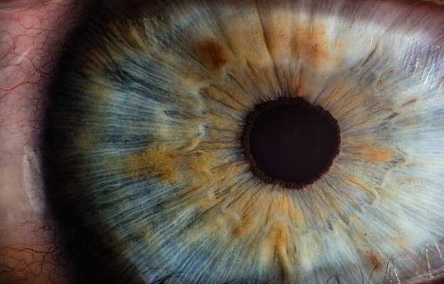
The eye can be compared to a digital camera. Much like a camera lens, the curved front surface of the eyeball, called the cornea, allows light to be focused. The iris, the coloured part of the eye, functions as a shutter or diaphragm and adjusts the pupil (black circular centre in the coloured iris which is like the aperture) size to regulate the amount of light entering the eye in order to capture the image. Through the pupil, light falls on the crystalline lens of the eye which focuses it more. The light focused by the cornea and the crystalline lens then falls on the retina (innermost layer of the eyeball) which acts as the image sensor and converts the optical image to an electronic signal and the optic nerve takes this signal to the visual cortex in the brain where it’s interpreted.

Eye cancer is an uncommon disease. Primary eye cancer, which begins in the eye, is rarer than secondary eye cancer which spreads from another body part to the eye. While the most common kind of eye cancer in adults is melanoma of the iris, ciliary body and choroid, only five out of a million people get diagnosed with it each year.

Broadly, there are two types of eye cancer. Those that occur in the structures within the eyeball (intraocular) and those that arise in the surrounding supportive tissues like eyelids. Often, eye cancer goes undiagnosed because it is generally painless. Deterioration of eyesight is the usual symptom. External signs may include bulging of the eyeball (proptosis) or lumps in surrounding structures.

Eye cancer is diagnosed using a mix of ophthalmoscopic examination, blood tests and imaging techniques. Definitive treatment could be surgical or radiotherapy. Laser therapy might be added for improved results. Prognosis is generally good unless the cancer has spread to other body parts.

What is eye cancer?

An uncontrolled growth of cells that originates and grows inside the eye is termed as eye cancer. These cells differ from the normal cells as their multiplication is uncontrolled and, hence, they’re termed atypical cells. In some cases, they may even extend beyond the eye to other parts of the body. The reverse may also occur; that is, atypical cells from tumours of other parts of the body may get deposited in or infiltrate the eye.

Types of eye cancers

Depending on where the eye cancer originates and how it spreads, it can be of different types:

  • Ocular melanoma: This is the most common type of eye cancer in the adult age group. Melanomas originate from the cells involved in the formation of pigments for different parts of the body including our eyes. This malignancy starts in the choroid of the eye, which consists of these pigmented cells. Uveal melanoma is said to be the most common form of malignant, primary intraocular eye cancer in grown-ups.
  • Primary intraocular lymphoma: A type of malignancy involving the white blood cells known as lymphocytes. It usually occurs in old immunocompromised patients like those suffering from HIV/AIDS.
  • Retinoblastoma: The most commonly occurring ocular malignancy in children, the pathology behind retinoblastoma is an inherited genetic mutation. This tumour starts in the retina, later on spreading to different parts of the eye and other organ systems.
  • Ocular metastasis: Sometimes different tumours that originate from other parts of the body, like lung cancer, can affect the eye. The malignant cells travel to the eye via blood vessels. The spread of cancer through blood is called a haematogenous spread.

Other eye tumours

Depending on where they occur, some other types of eye tumours are:

Choroidal tumors: The choroid is a vascular layer in the eye that provides oxygen and nutrients to all parts of the eye, including the retina.

  • Choroidal hemangioma: These tumours are benign but may cause eye damage.
  • Choroidal melanoma
  • Choroidal metastasis: Cancer can metastasize from other parts of the body to reach the eye. Breast cancer in women and lung cancer in men are the most common causes, though the cancer could also originate in the prostate, kidneys, thyroid or gastrointestinal tract.
  • Choroidal osteoma: These are benign but ossifying tumours in which the vascular layer of the eye (choroid) is replaced by a bony growth.
  • Ciliary body melanoma: This is a type of uveal melanoma. The ciliary body exists around the iris and includes the ciliary muscle which helps to con

Conjunctival tumours 

  • Kaposi sarcoma: This is a vascular neoplasm (tumour) usually associated with the conjunctiva and eyelids.
  • Epibulbar dermoid: These are benign yellowish or pinkish tumours that may cover the cornea and the whites of the eyes (sclera).
  • Malignant conjunctival tumors
  • Lymphoma of the conjunctiva
  • Melanoma and PAM with atypia
  • Pigmented conjunctival tumors
  • Pterygium (surfers’ eye)
  • Squamous carcinoma and intraepithelial neoplasia of the conjunctiva

Eyelid tumours

  • Kaposi sarcoma
  • Basal cell carcinoma
  • Squamous cell carcinoma
  • Sebaceous cell carcinoma
  • Malignant melanoma

This is not an exhaustive list of tumours that can affect the eyes, but only a list of the more common conditions. Please also note that not all these tumours are cancerous, some are benign.

Symptoms of eye cancer

Ocular malignancy may or may not be symptomatic and hence may only be detected during a routine eye checkup.

Common symptoms include: 

  • Painless loss of vision (most common)
  • Shadows or light flashes
  • Blurring of vision
  • Progressively increasing dark patches in vision
  • Bulging of eye or proptosis 
  • Lump in eye or on the eyelid; it may increase in size 
  • Eye cancer can rarely present as ocular (eye) pain

Risk factors of eye cancer

There are numerous factors that can increase the risk of developing eye cancer. They are as follows: 

  • Age: Eye cancer most commonly affects people aged 50-60 years. It is rare above the age of 70 years.
  • Race: Caucasians have a higher tendency to develop melanomas than Black people. 
  • Certain eye conditions: People with various conditions such as:
    • Pigmentation of the eye or surrounding skin (melanocytosis) 
    • Moles in the eye
  • Genetic transmission: Some eye cancers may be inherited genetically such as retinoblastoma.  
  • Environmental factors: Some studies suggest excessive sunlight exposure may also contribute to the development of eye cancers.

Prevention of eye cancer

Eye cancer in general is not regarded as entirely preventable as not many modifiable risk factors exist. Therefore, limiting the most significant factor, direct sunlight and UV exposure, are key to primary prevention.

Primary prevention

  • Family history: Being aware of pre-existing eye cancer in the family is essential for early detection. One out of two children born to a parent with heritable retinoblastoma is likely to suffer from it as well.
  • Reducing sunlight exposure: Much like melanomas of the skin, melanomas of the eye are also linked to prolonged exposure to UV rays. Limiting the time you spend in the sun is the best precaution.
  • Sunglasses: The American Cancer Society recommends sunglasses with 99-100% UV-A and UV-B absorption as protection when you go out in the sun. Wraparound styles offer the most protection to the eye and surrounding skin.

Secondary prevention: Secondary prevention, or early detection and effective treatment, are the most important when it comes to eye cancer.

  • Eye examination at birth: Retinoblastoma, the most common eye cancer afflicting children, can be detected at birth in some cases. The most common sign to look out for is leukocoria, or white pupil. This means that the central black portion in the coloured part of the eye is white instead of the usual black.
  • Eye checkups: It is recommended that healthy adults between 18 to 40 years of age without any risk factors or family history of eye disease get a check-up every two to three years. For those who are 41 to 60 years old, a visit to the eye doctor every two years is beneficial. Those above 60 or with any other risk factors should get their eyes tested annually.

Diagnosis of eye cancer

If a general physician suspects eye cancer during the patient’s check-up he/she should refer the patient to an ophthalmologist.

An ophthalmologist will perform a thorough physical examination and numerous investigations to confirm the diagnosis.

  • Eye examination: Instruments such as ophthalmoscopes are utilised to examine the different parts of the eye in detail.
  • Ultrasound scan: The doctor will place a probe over the patient’s eye, which will generate high frequency sound waves and develop a picture of the interior parts of the eye (ultrasound), aiding the doctor in localising the tumour and measuring its size. 
  • Fluorescein angiography: A fluorescent dye is injected in the blood which travels throughout the body and reaches the blood vessels of the eye. The ophthalmologist will take many pictures of the eye using a special camera to rule out any other diseases affecting the eye.
  • Fine needle biopsy: This is rarely used as most ocular tumours can be identified without a biopsy. Procedure involves removing some cells using a fine needle and assaying them microscopically.
  • Genetic testing: Helps provide information about the likelihood of recovery, or prognosis, and formulating a treatment plan for the patient by detecting tumour-specific genes and proteins. Testing is performed on tissue sample obtained during biopsy or surgical excision.
  • Testing for spread of tumour: Most common site of metastasis of eye cancer is the liver. To check for liver metastasis, a liver function test is ordered and this may be supplemented by radiological investigations such as an ultrasound or a computed tomography scan (CT scan) of the abdomen.
  • CT scan: Generates 3D images using an X-ray machine. Allows doctors to measure the size of the tumour and accurately identify the site.
  • Magnetic resonance imaging (MRI): Magnetic resonance imaging (MRI) performs the same function as a CT scan—that is measurement and localisation of tumour and detecting any metastasis. The major difference is that MRI uses magnetic waves instead of X-rays.
  • Positron emission tomography (PET) scan:positron emission tomography (PET) scan is based on the principle that cancer cells utilise more sugar (glucose) than normal cells. Hence, radioactive labelled glucose is injected in the patient and its uptake by atypical cells is measured. A scanner detects this and helps in the generation of images of the organ involved.

Staging of eye cancer

The TNM system is a method to describe the stage of a particular tumour. 

It consists of three parts: 

  • Tumour (T): Size and location of the tumour 
  • Node (N): Whether the tumour has spread to any lymph nodes 
  • Metastasis (M): Whether the cancer has metastasised to any other part of the body

After evaluating each part of the TNM system, the results are combined and the tumour is staged. There are a total of four stages. The stage of the cancer helps in deciding the most appropriate treatment plan for the patient.

A simplified version of staging of eye cancers is as follows:  

  • Stage 1: The tumour is small (less than 3mm)  and does not involve any other parts of the eye or any other organ system.
  • Stage 2: Tumour size slightly larger than stage 1 and may or may not have spread to other eye parts or other organ systems. 
  • Stage 3: Large tumour size and spread to other parts of the eye.
  • Stage 4: Cancer has spread to other parts of the body (Metastasis)

Treatment of eye cancer

The best approach to the treatment of any kind of cancer is with a multidisciplinary team. This team usually consists of various specialists who are involved in different aspects of patient care and treatment.

Apart from ophthalmologists and oncologists, physician assistants, plastic surgeons, radiologists, nurses, social workers and dieticians are essential when making decisions regarding patient care. 

The basic concept behind the treatment protocol for any type of cancer apart from curing cancer itself is to prevent its spread to other regions in the body, to maintain the quality of life and to minimise any limitations that cancer and its treatment will cause.

The various treatment protocols that are offered as per established guidelines are as follows:

  • Active surveillance: This involves closely monitoring the patient. This approach is followed for patients with a tumour that is small, non-aggressive or in cases where the benefits of subjecting the patient to medical treatment are outweighed by the risks or rendered moot by a poor prognosis. At times, end-of-life care, or palliative care, is more important than aggressive therapy. If the cancer shows signs of rapid growth (more than 10mm diameter) or spread to other organs (most common being the liver), this “wait and watch” protocol can be modified to active treatment.
  • Surgery: Is done in order to remove the tumour along with some healthy surrounding structures, this is known as tumour resection. It is one of the most common treatment modalities for eye cancer. The amount of tissue removed depends on the stage of cancer.
    The surgeon may perform:
    • Iridectomy or removal of part of iris (structure controlling size of pupil)
    • Iridocyclectomy: Removal of part of iris and ciliary body (structure controlling shape of lens) 
    • Enucleation: Removal of whole eye
  • Radiotherapy: High energy X-rays are utilised to destroy malignant cells. Radiotherapy is performed by a radiation oncologist. The doctor may use external beam radiation using a machine outside the body or may use implants (radioactive discs placed near the tumour) for internal radiation therapy.
  • Laser treatment: The heat generated from a laser works by reducing the size of tumour making it easier to remove surgically or treat with radiation therapy. The treatment has fewer side effects and is thus advantageous.

Side effects of various treatment protocols for eye cancer

The following are some of the key side effects of cancer treatments for the eye:

Surgery side effects

  • Infection at surgical site 
  • Effects of anaesthesia (headachedizziness)
  • Pain
  • Recurrence of tumour
  • Reduction or loss of vision

Radiotherapy side effects

  • Cataract: clouding of lens of the eye. It causes reduction of vision and glare from sunlight.
  • Loss of eyelashes
  • Dryness of eye

Follow up:

To minimise the side effects of treatment, ensure the patient’s well-being and catch any recurrence, a comprehensive follow-up plan must be drawn up after completing active therapy.

Follow-ups usually schedule a doctor’s visit every 6-12 months and generally consist of physical examination, blood tests and radiological imaging which could be X-ray, ultrasound, CT scan or PET scan.

Eye cancer prognosis or chances of survival

Prognosis in this case is measured in terms of five-year survival rate which is based on the percentage of people who survive for at least five years after the diagnosis of cancer is made.

Overall for eye cancers, this number is around 80% and if diagnosed earlier, this percentage increases to 85%.

If the tumour has spread to surrounding organs or lymphatics then the five-year survival rate drops to 71%.

In case of distant spread to other organ systems, this number further reduces to 13%.


References

  1. Pe'er J. Pathology of eyelid tumors. Indian Journal of Ophthalmology, March 2016; 64(3): 177-90. PMID: 27146927.
  2. Ocular Melanoma Foundation US [internet]. Types of eye cancer.
  3. Singh A.D., Turell M.E. and Topham A.K. Uveal melanoma: trends in incidence, treatment, and survival. Ophthalmology, September 2011;118:1664–78.
  4. R.C Eagle, Jr. The pathology of ocular cancer. Eye (Lond), February 2013; 27(2): 128–136. Published online 2012 Nov 16.
  5. P. De Potter. Ocular manifestations of cancer. Current Opinion in Ophthalmology, December 1998; 9(6): 100-4. PMID: 10387328.

Medicines for Eye cancer

Medicines listed below are available for Eye cancer. Please note that you should not take any medicines without doctor consultation. Taking any medicine without doctor's consultation can cause serious problems.