What is Two-dimensional Echocardiography?

Two-dimensional echocardiography (2D ECHO) is a test that enables a physician to visualise the beating heart through the simple use of ultrasound waves generated from a handheld device known as a transducer. A transducer contains a special type of a crystal known as a piezoelectric crystal (similar to quartz), which produces sound waves when varying voltage is applied to it. The produced sound waves reflect off different tissues of heart towards which the device is pointed. These waves are captured back by the crystal in the receiving mode and converted to electrical signals, which can be used to form an image of the heart on a monitor.

There are different types of 2D ECHO tests, and transthoracic echocardiography (TTE) is the standard type among these. TTE is based on the principle that ultrasound waves, (i.e., waves of a frequency greater than 20 kHz inaudible to the human ear) are poorly transmitted through bone and air but are reflected at the boundaries between two tissues. Thus, by holding the transducer at various locations on the chest, known as acoustic windows, a 2D image of the heart from various angles can be generated.

  1. Why is 2D ECHO performed?
  2. How is 2D ECHO performed?
  3. How do you prepare for 2D ECHO?
  4. What do 2D ECHO results indicate?

2D ECHO tests are performed mainly to detect various heart diseases and problems. A doctor would recommend 2D ECHO tests to gain a better understanding of the following:

  • Size and shape of the heart and its chambers: Heart may enlarge in size due to high blood pressure (BP), or the lower chambers of the heart, the ventricles, may enlarge due to different causes and lead to inefficient pumping of blood.
  • Pericardium, i.e., the sac that surrounds the heart: In certain cases, a fluid build-up may occur inside the pericardium and cause compression of heart leading to signs and symptoms, such as a drastic fall in BP, quiet heart sounds, shortness of breath, weakness and cough.
  • Heart muscles: Heart attacks may lead to weakening of heart muscles, which further decreases blood supply to the heart.
  • Heart valves: A 2D ECHO test can be used to correlate the symptoms of malfunctioning heart valves, such as fatigue, leg swelling and changes in heart sounds on auscultation
  • Blood clots: Patients with ongoing or a history of stroke are recommended this test to detect the location and extent of heart tissue damage.
  • Congenital heart defects: It is possible to detect heart problems in infants, such as a hole in the heart wall, through 2D ECHO.
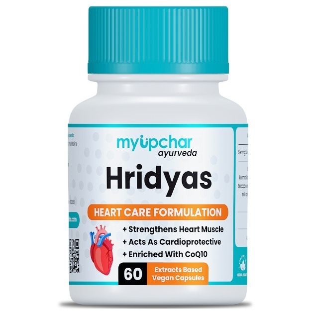
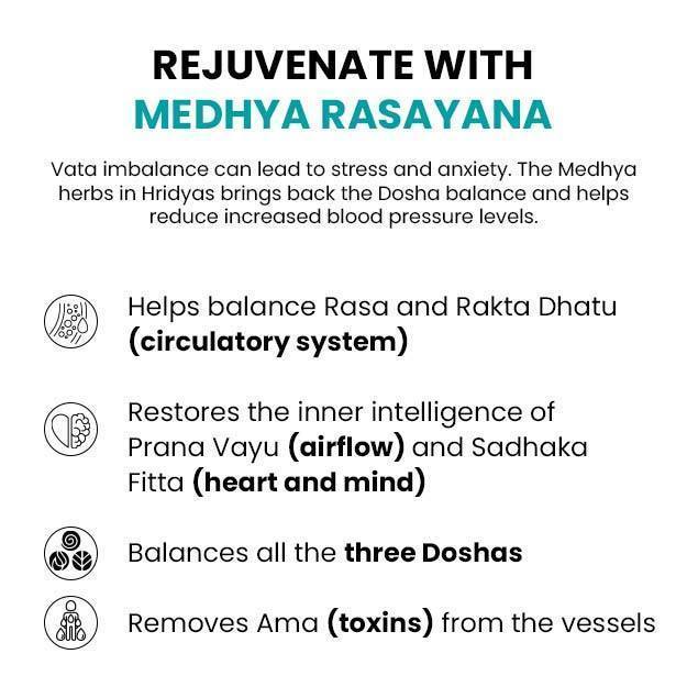
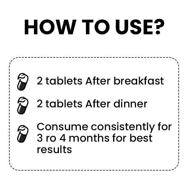
myUpchar doctors after many years of research have created myUpchar Ayurveda Hridyas Capsule by using 100% original and pure herbs of Ayurveda. This Ayurvedic medicine has been recommended by our doctors to lakhs of people for problems like high blood pressure and high cholesterol, with good results.
BP Tablet
₹899  ₹999  10% OFF
BUY NOW

A 2D ECHO test is performed with a handheld transducer unit that is portable to patients’ bedside. The procedure for this test depends on the type of test being performed.

  • Transthoracic echocardiography: In this procedure, transducer head, which emits ultrasound waves, is placed on the chest at various positions to get instantaneous images of the heart.
  • Stress echocardiography: It is conducted in two phases. In the first phase, a resting echocardiogram is taken where the individual is made to lie down on the left side with their left arm stretched out. In the second phase, the individual will be made to perform moderate exercises, such as walking on a treadmill on an incline for 5-15 minutes depending on their fitness level and age. During this phase, their BP and electrocardiogram will be constantly monitored using a BP cuff placed on the upper arm and electrodes placed on the chest and legs. Seldom, individuals may experience discomfort in chest, dizziness and headache during this procedure. Drugs, such as sildenafil, can affect the results of this test.
  • Transoesophageal echocardiography: In this procedure, the transducer device is attached to a probe, which is inserted into the throat and emits ultrasound waves that reflect from the heart, to obtain a different angle of view of the heart. The individual may be injected with medicine to help him/her relax during the test, and the back of the mouth will be numbed with a spray or gel prior to inserting the probe. An individual may experience a sore throat after the completion of this test and must only resume eating and drinking 30-60 minutes after the test.
  • For TTE, no specific preparation is required prior to the test, making it easier and more advantageous than other tests when it comes to preparation.
  • For stress echocardiography, the individual must not eat or drink anything for three hours before the test. Loose and comfortable clothing must be worn. Also, it is important to inform the physician if you are taking medicines such as sildenafil (Viagra).
  • TEE is mostly done for hospitalised patients. Fasting for several hours is required before this test, and the exact duration for fasting is informed by the treating physician. The patient must inform the physician about allergies to any medications. Dentures must be removed prior to the test.
Spirulina Capsules
₹539  ₹599  10% OFF
BUY NOW

A normal test result reflects normal functioning; structure; and movement of heart muscles, valves and chambers.

An abnormal 2D ECHO (both TTE and TEE) test could indicate a myriad of heart problems. It requires consultation with a cardiologist for a final diagnosis and treatment.

A normal stress echocardiogram reveals that the heart is able to function normally even under stress. Any abnormality in stress echocardiography reveals decreased blood flow to a particular part of the heart due to blockage or narrowing of blood vessels supplying that part.

Disclaimer: All results must be clinically correlated with the patient’s complaints to make a complete and accurate diagnosis. This information is purely from an educational perspective and is in no way a substitute for medical advice from a qualified doctor.

References

  1. Alaa A. Mohamed, Ahmed A. Arifi, Ahmed Omran. The basics of echocardiography. Journal of the Saudi Heart Association (2010) 22:71–76.
  2. Rick A. Nishimura, Raymond J. Gibbons, James F. Glockner, A. Jamil Tajik. Harrison’s Principles of Internal medicine. Noninvasive Cardiac Imaging: Echocardiography, Nuclear Cardiology, And MRI/CT Imaging.. 16th edition. 1320.
  3. Spodick, DH (Aug 14, 2003). Acute cardiac tamponade. The New England Journal of Medicine (2003) 349(7): 684–690.
  4. Fihn SD, Blankenship JC, Alexander KP, Bittl JA, Byrne JG, Fletcher BJ, Fonarow GC, Lange RA, Levine GN, Maddox TM, Naidu SS, Ohman EM, Smith PK. 2014 ACC/AHA/AATS/PCNA/SCAI/STS focused update of the guideline for the diagnosis and management of patients with stable ischemic heart disease: a report of the American College of Cardiology/American Heart Association Task Force on Practice Guidelines, a. J Am Coll Cardiol. 2014 Nov 4;64(18):1929-49. PMID: 25077860
  5. Boden WE. Angina pectoris and stable ischemic heart disease. In: Goldman L, Schafer AI, eds. Goldman-Cecil Medicine. 25th ed. Philadelphia, PA: Elsevier Saunders; 2016:chap 71.
  6. Solomon SD, Wu JC, Gillam L, Bulwer B. Echocardiography. In: Zipes DP, Libby P, Bonow RO, Mann DL, Tomaselli GF, Braunwald E, eds. Braunwald's Heart Disease: A Textbook of Cardiovascular Medicine. 11th ed. Philadelphia, PA: Elsevier; 2019:chap 14.
  7. National Heart, Lung, and Blood Institute [Internet]. Bethesda (MD): U.S. Department of Health and Human Services; Transesophageal Echocardiography

Related Articles

Upper GI Endoscopy

Dr. Ayush Pandey
MBBS,PG Diploma
7 Years of Experience

Obesity Profile

Dr. Ayush Pandey
MBBS,PG Diploma
7 Years of Experience

Extractable Nuclear Antigen ...

Dr. Ayush Pandey
MBBS,PG Diploma
7 Years of Experience
Read on app
cross
Ask your health query from live doctors now!