Macular Edema

Dr. Suvansh Raj NirulaMBBS

February 22, 2021

January 29, 2024

Macular Edema
Macular Edema

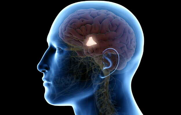
The eyes process visual input as images that are transmitted to the brain by light rays falling on a thin layer over the back surface called the retina. The retina consists of specialised cells, rods and cones that process shapes and colours of images. Within the retinal layer is a central area called the macula, which helps sharpen and clarify the image being viewed. The fovea is a centralised pit or depression in the macula of the retina that provides the greatest visual acuity.

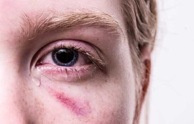
Certain diseases that affect the eye can cause fluid and protein to accumulate and deposit in the macula of the retina, thus producing macular edema. It is a common final stage of many ocular diseases (conditions pertaining to the eyes). Common causes of macular edema include diabetic retinopathy (diabetes mellitus induced eye disease), occlusion of blood vessels within the eye (for example central retinal vein occlusion and branch retinal vein occlusion), chronic uveitis and age related macular degeneration (AMD). Other important causes are due to optic lens insertion after cataract surgery and with certain medications.

Breakdown of the blood retinal barrier (BRB) due to inflammatory disease processes and increased vascular permeability causes fluid and proteins to abnormally cross over into the macula and deposit there. The severity of macular edema depends one the extent of the area affected, the distribution of the swelling (focal or diffuse) and its proximity to the fovea centralis, which determines the overall visual acuity. Retinal thickness and cysts due to edema and traction on the vitreous humour also determine the severity of macular edema. Broadly, two types of macular edema are described – cystoid macular edema or diabetic macular edema. Certain types of age related macular degeneration (AMD), “wet” or educative AMD, can be considered as a third classification but technically they also ultimately lead to cystoid macular edema.

(Read more: Eye disorders)

Types of macular edema

Following are the two types of macular edema:

  • Cystoid macular edema: Fluid and protein accumulates in the outermost layer of the macula, called the plexiform layer, and cysts (hence called cystoid) form in the retina. The blood-retinal barrier (BRB) breaks down and clear fluid-filled cysts form in the retina, displacing the retinal photoreceptor cells. Although it usually resolves in a few months, if it persists for a prolonged period the photoreceptors, and thus vision, may be permanently lost. Cystoid macular edema is the resultant end-stage of many eye diseases such as central retinal vein occlusion (CRVO), branch retinal vein occlusion (BRVO), Irvine-Gass syndrome, some eye drop drugs and ocular inflammatory diseases.
  • Diabetic macular edema: Long-standing raised blood glucose levels (hyperglycaemia) in diabetes mellitus cause certain changes in the blood vessels and capillaries of the body (including the eyeball), causing fluid to leak out from them. This results in fluid and protein accumulation in the macula. Oxidative stress and certain factors (like VEGF) that cause fibrosis and thickening of the area of edema are also produced. Diabetic retinopathy (which can be of two types: proliferative and non-proliferation) progresses to diabetic macular edema, which can be focal or diffuse. End-stage diabetic eye disease (EDED) follows diabetic macular edema and can present with vitreous hemorrhage and retinal detachment.

Signs and symptoms of macular edema

Although the presentation varies with the underlying cause, usually the patient presents with painless and gradual impairment, or total loss, of central vision. Some signs and symptoms affecting the patient’s vision can include:

  • There may be a scotoma (a black spot within the field of vision).
  • Visual acuity (ability to discern different shapes and details of the objects being viewed) is impaired. It can typically lie in the region of 6/12 to 6/60 on testing with Snellen’s chart.
  • Patients with Irvine-Gass syndrome (clinical term for macular edema occurring due to optic lens insertion following cataract surgery) usually experience a gradual blurring, with good initial vision.
  • In the case of age-related macular degeneration (AMD), patients may complain of visual distortion (metamorphopsia), particularly of straight lines.
  • Sometimes, blue-yellow colour blindness can set in.
  • There may be a loss of contrast sensitivity.
  • In some cases, colours in the central vision appear 'washed out' or desaturated.
  • Sometimes, clinically significant macular edema may be asymptomatic, especially in the early stages, and is only detected upon ophthalmological eye examination. Therefore, it is recommended that patients with diabetes mellitus get their eyes tested frequently.

(Read more: Vision problems in babies)

Causes of macular edema

Following are some of the causes of macular edema:

  • Diabetes mellitus and diabetic retinopathy 
  • Age related macular degeneration (AMD)
  • Vitreous traction
  • Retinal vein occlusions:
    • Central retinal vein occlusion (CRVO)
    • Branch retinal vein occlusion (BRVO)
  • Inherited genetic disorders:
  • Inflammatory eye diseases:
    • Uveitis
  • Autoimmune diseases:
    • Behçet syndrome
  • Post cataract surgery: Optic lens insertion following cataract surgery causes macular edema known as Irvine-Gass syndrome.
  • Certain medications like latanoprost and timolol.
  • Eye tumours 
  • Trauma

Risk factors of macular edema

Patients with diabetes mellitus (type 1 and type 2) are at an added risk of diabetic macular edema in case of:

Diagnosis of macular edema

Following steps are taken to diagnose macular edema:

  • History: The doctor begins by taking a detailed and thorough medical history of the patient. Keen attention is paid to complaints pertaining to deterioration of vision (current status and progression), systemic signs of undiagnosed diabetes mellitus (increased thirst, increased urination, increased hunger, involuntary weight loss, tingling or loss of sensation in hands or feet, unexplained fainting spells, etc.) and a history of preexisting eye disease. A history of ocular diseases that may be running in the family and medication history is also noted. Following history taking, a comprehensive physical examination is conducted, which includes specialised ophthalmological eye tests.
  • Physical examination: Following a medical history, a physical examination of the patient is done by the doctor. Signs of systemic diseases that produce macular edema can be elicited. Blood pressure is also taken, as hypertension can produce retinal vein occlusion diseases. A neurological examination is also important to rule out some other possible causes of visual deterioration. An ophthalmological examination is paramount.
  • Ophthalmological examination (eye testing): The ophthalmologist (eye doctor) conducts an eye exam, geared at eliciting the deficits in vision, the components of which are:
    • Visual acuity testing: The patient is made to stand six metres away from a Snellen’s chart and is made to read out alphabets and numbers on it. If an individual can read the smallest letters from a distance of 6 metres, they are said to have 6/6 perfect vision.
    • Ishihara test: Colour blindness can be diagnosed using specialised charts with digits written in coloured dots. If the patient is able to make out the colours and read the digit their colour vision is functioning.
    • Amsler grid test: This is another chart that consists of vertical and horizontal lines and is useful in assessing the patient’s central vision. The lines may appear bent in case of macular disease.
    • Ophthalmoscopy: A test that allows the doctor to see signs of eye diseases inside the eye.
    • Retinoscopy: A special test to diagnose and grade refractive errors of the eyes that may be affecting vision.

(Read more: Swollen eyes)

If you are tired of dieting and exercising and are not able to lose weight, then use myUpchar Ayurveda Medarodh Fat Burner Capsule, it has no side effects, order it today and avail the benefits.

Differential diagnosis of macular edema

Many different underlying diseases, both systemic and of the eye particularly, can cause macular edema. It is important to diagnose them early and to classify them accurately to help guide treatment, overall management and outcome. However, some other ocular diseases can also present with gradual painless visual impairment or loss and need to be considered while arriving at a diagnosis. Other causes of gradual painless impairment or loss of vision include:

  • Refractive errors (nearsightedness, farsightedness, age related diminution of vision)
  • Progressive pterygium of the eye (Read more: Double vision)
  • Senile cataract
  • Developmental cataract
  • Chronic Simple Glaucoma
  • Corneal dystrophy
  • Corneal degeneration
  • Retinal dystrophy
  • Optic nerve atrophy

Tests for diagnosing macular edema

Following are the tests required to diagnose macular edema:

  • Blood tests:
    • Blood glucose levels: Uncontrolled diabetes mellitus is the most common cause of macular edema (diabetic macular edema).
    • Kidney function test: Kidney disease may be lined with worsening of eye disease in some cases like diabetes mellitus.
    • Blood lipid panel: Raised blood cholesterol and lipids can accelerate diabetes eye disease.
    • Immunological tests: Autoimmune diseases like Behçet syndrome can be diagnosed.
  • Urine tests:
    • Urine ketone and glucose: Uncontrolled diabetes mellitus can be detected.
  • Ophthalmological imaging studies:
    • Fluorescein angiography: A special dye called fluorescein is injected through the patient’s arm and it travels to the eyes quickly. A camera takes pictures of the retina and the vessels in it. Fluid leakage from blood vessels in the retina is picked up. 
    • Optical coherence tomography (OCT): A type of specialised imaging that takes pictures of the retina and can pick up very minute changes. 
myUpchar doctors after many years of research have created myUpchar Ayurveda Urjas Energy & Power Capsule by using 100% original and pure herbs of Ayurveda. This Ayurvedic medicine has been recommended by our doctors to lakhs of people for problems like physical and sexual weakness and fatigue, with good results.
Power Capsule For Men
₹719  ₹799  10% OFF
BUY NOW

Management of macular edema

Management of diabetic macular edema is similar to cystoid macular edema but also focuses on strictly controlling blood glucose levels. Management of macular edema depends on the underlying cause; for example, uveitis or retinitis needs specific medical treatment. Some of the methods used to treat cystoid macular edema are described here:

(Read more: Self-monitoring of blood glucose)

Medical management:

  • NSAIDs: Topical eye drops or oral NSAIDs after cataract surgery can reduce inflammation and prevent macular edema.
  • Corticosteroids: They can be used as eye drops, oral drugs or intravitreal injections to reduce inflammation. Intravitreal triamcinolone injections are useful in uveitis induced macular edema.
  • Carbonic anhydride inhibitors: These drugs act on molecular ion channels and clears excessive fluid and can be beneficial.
  • Anti-VEGF injections: Intravitreal injections of anti-VEGF agents like Bevacizumab and Ranibizumab can stop the action of VEGF that produces fibrosis and thickening of the macula.

Surgical management:

  • Laser pharmacotherapy: The dead and diseased portion of the photoreceptor rich pigmented retinal epithelium is removed by heat energy from a light source. This allows more oxygen and blood supply to be received by the healthy photoreceptor cells.
  • Vitreolysis: Certain drug agents that can be injected into the vitreous humour that has become dense and thickened by inflammation due to macular edema. These agents dissolve the fibrous thickening.
  • Vitrectomy: If medical therapy fails, the thickened and inflamed portion of the vitreous humour may be surgically removed.

Complications of macular edema

Following are some of the complications that can occur due to macular edema:

  • Advanced diabetes eye disease
  • Retinal detachment
  • Retinal hole
  • Vitreous haemorrhage
  • Irreversible loss of vision

Prevention of macular edema

Following are the steps you can take to prevent macular edema:

  • Controlling blood glucose levels in diabetes mellitus (both type 1 and 2)
  • Correcting lifestyle diseases like hypertension and raised cholesterol levels
  • Annual ophthalmological examination (eye tests) are recommended in all diabetics to detect asymptomatic retinopathy
  • Adherence to postoperative medications like NSAIDs after cataract surgery

Prognosis of macular edema

Although outcomes depend on the underlying cause of macular edema, cystoid macular edema has a better prognosis. It generally resolves spontaneously within a few months; however, surgical management may be necessary. If irreparable damage hasn’t occurred, vision may return to normal. Diabetic macular edema is generally progressive and causes extensive damage and usually leads to permanent visual impairment.

(Read more: Blindness)