Download App


Lithotripsy literally translates to ‘breaking of stones’. It is a medical procedure that is typically non-invasive and is used to break stones in the gallbladder or kidneys with ultrasonic or laser energy. Bezoars can be treated this way as well. Lithotripsy is most commonly used to treat large kidney stones (greater than 5 millimetres in dimension) that cannot be passed in the urine with increased fluid intake. Newer applications of lithotripsy, like that as second-line treatment of soft tissue injuries, are also available today. Many other stones in the body can be treated with lithotripsy.
- Gallstones (Cholelithiasis)
- Kidney stones (Nephrolithiasis)
- Bezoars
- Types of lithotripsy
- Indications of lithotripsy
- Preparation for lithotripsy
- Contraindications of lithotripsy
- Complications of lithotripsy
- Prognosis of lithotripsy

Gallstones (Cholelithiasis)
The gallbladder is a small pouch-like organ present on the underside of the liver. The primary function of the gallbladder is to hold bile, a green enzymatic fluid that helps in the digestion of certain foods (mostly fats), and to secrete mucus. Sometimes, due to supersaturation of the bile with cholesterol or hemoglobin (the red oxygen-carrying pigment of blood) breakdown products due to varying underlying diseases, crystallisation may occur and stones may form. These stones can form in any part of the biliary tract (gallbladder or bile ducts) and can also migrate to other parts of the system. While most gallstones remain asymptomatic, inflammation of the gallbladder (cholecystitis) or bile duct (cholangitis) can produce various symptoms including – abdominal colic (pain), jaundice, fever and more. Three types of gallstones can form:
- Cholesterol stones: Cholesterol stones are the most common type of gallstones and are yellow in appearance and generally solitary. These stones are composed of more than 80% cholesterol and generally occur due to poorly-controlled blood cholesterol levels (hypercholesterolaemia), which leads to supersaturation of bile and stones to form.
- Pigment stones: Pigment stones are typically dark-coloured, brown to black, and are smaller and numerous. They contain less than 20% of cholesterol and are composed mainly of the blood hemoglobin metabolic breakdown product bilirubin. The pigment bilirubin gives it a dark colour. Typical causes of pigment stones include undue haemolysis (excessive breakdown of red blood cells). which could be caused by various acquired and inherited diseases.
- Mixed stones: Stones that are composed of both pigment bilirubin, as well as 20-80% of cholesterol, are called mixed stones. They can be yellow-brownish in colour.

Kidney stones (Nephrolithiasis)
Kidneys are organs that filter the blood of toxic waste material and create urine to clear it. Sometimes salts and minerals of the urine can supersaturate within the kidneys and stones may form. While many small stones can simply be passed in urine and treated by drinking plenty of fluids, bigger stones (generally larger than 5 millimetres in dimension) can become symptomatic and painful, requiring external methods of extraction. At times, kidney stones can lead to other complications like infections of the urinary tract. Stones may form in, or migrate to, the ureters (tubes connecting the kidneys to the urinary bladder) as well. Typically, five types of kidney stones, based on their constituent minerals, are known to form:
- Calcium oxalate stones: Calcium stones are the most common stones to form in the urinary tract. Oxalate is derived from a person’s diet and excessive consumption of certain foods can lead to its accumulation. Other causes may also lead to calcium oxalate kidney stones.
- Calcium phosphate stones: Another type of calcium kidney stone that occurs is made of calcium phosphate. Phosphate stones are generally linked to metabolic disorders. Some medications can also raise phosphate levels, leading to supersaturation of urine and ultimately stone formation.
- Struvite stones: pH alterations (acid-base balance) due to bacterial urinary tract infections can cause magnesium amino phosphate stones to form. These stones can grow to big sizes relatively quickly.
- Uric acid stones: Hyperuricaemia (raised serum levels of uric acid) can precipitate the formation of urate stones. A high protein diet, use of certain medications, genetic factors, lifestyle diseases like diabetes mellitus and gout are all possible factors that cause uric acid stones.
- Cystine stones: These stones are generally resultant of a genetic disorder called cystinuria in which the non-essential amino acid cysteine is not metabolised adequately and excessive metabolite, called cystine, is secreted in urine.
(Read more: Kidney stone diet)
Bezoars
Sometimes, undigested or partially digested material may accumulate in the digestive tract and form tightly-packed hardened masses called bezoars. Bezoars can occur in people of all ages and are most commonly found in the stomach. Various kinds of bezoars can form including:
- Phytobezoars: These bezoars are the most common kind and are usually made up of indigestible vegetable fibres.
- Diospyrobezoars: Unripened persimmons fruit consumption leads to diospyrobezoar formation.
- Trichobezoars: People who suffer from trichophagia or the psychiatric disorder that urges one to consume hair can have large bezoars form of hair and food in their digestive tracts.
- Pharmacobezoars: Undissolved medication can form semi-solid masses in the alimentary tract. These bezoars are typically seen with overdose or overconsumption of sustained-release medicines.
- Lactobezoars: Bezoars composed of milk protein and mucus can form in premature infants being fed formula milk.
- Foreign body bezoars: Accidental swallowing of foreign bodies by children can lead to bezoar formation.
- Faecolith: The accumulation of solidified and impacted faeces in the large intestine is called a faecolith.
Types of lithotripsy
The principle of lithotripsy is to break stones in the body using sound wave or laser energy in a non-invasive manner as an alternative to surgical removal. Lithotripsy techniques can be categorised as:
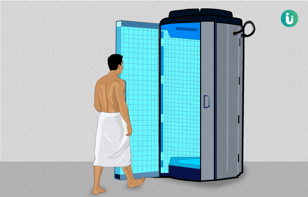
- Extracorporeal lithotripsy: The application of wave energy to break up stones from outside of the body. The best example of this type of lithotripsy is extracorporeal shock wave lithotripsy (ESWL).
- Extracorporeal Shock Wave Lithotripsy (ESWL): This is the most common and preferred mode of lithotripsy. It is mostly used for large kidney stones (greater than or 5 millimetres in dimension) that cannot be passed in urine naturally and gallstones. The patient is sedated or anaesthetised before the procedure in order to help them stay still and not experience any anxiety. Afterwards, a specialised device called a lithotripter is applied externally to deliver a focused, high-intensity acoustic pulse that breaks up the stone in question. In the case of kidney stones, the stone fragments are then easily removed through the urine. The entire procedure takes up to an hour but can vary depending on the site, number or size of the stones. ESWL is most effective for stones smaller than 10 millimetres in dimension. The patient is generally discharged on the same day. ESWL has also come to be used for salivary stones (sialolithiasis) and as second-line therapy for soft tissue injuries like:
- Intracorporeal lithotripsy: While incisions are not needed, in this type of lithotripsy, scopes are inserted into the body for stone removal. Although other types of intracorporeal lithotripsy exist, the most common, effective and popular is the flexible ureteroscopy and laser lithotripsy (FURSL).
- Flexible Ureteroscopy and Laser Lithotripsy (FURSL): Also known as flexible ureteroscopy, lasertripsy is a type of lithotripsy used exclusively for stones of the urinary tract (kidneys, ureters, urinary bladder, or urethra). Flexible ureteroscopy and laser lithotripsy (FURSL) is preferred in cases where the stone is very large (greater than 2 centimetres/ 2 millimetres in size), irregularly shaped, hard to locate or causing bleeding. After placing the patient under general anaesthesia (GA), the procedure is started. A special scope with a camera (ureteroscope) is inserted into the urinary tract via the urethra and the impacted stone is located. After locating the stone, a fibre is passed through the scope that produces a laser beam to break up the stone. Fragments of the stone are collected by a basket in the scope and a temporary stent is placed in the ureter to improve urine flow and allow the removal of tinier stone fragments that may remain. The entire procedure, including the anaesthesia process, may last up to two hours. FURSL is preferred over extracorporeal shock wave lithotripsy (ESWL) in case the stone size is larger than 20 millimetres.
See Similar Category Medicines Here
Indications of lithotripsy
Lithotripsy is typically used to break up stones in the body. However, recently, some modalities of lithotripsy have come to be used in other applications, such as those of physiotherapy. Lithotripsy can be used in:
- Kidney stones
- Ureteric stones
- Bladder stones
- Urethral stones
- Gallstones
- Bile duct stones
- Bezoars
- Salivary gland stones
- Tennis elbow
- Achilles tendinitis
- Plantar fasciitis
- Shoulder rotator cuff injuries
Preparation for lithotripsy
The doctor will take a detailed medical history and conduct a thorough physical examination before setting a date for the procedure. Additionally, blood, urine and radiological tests may also be conducted. It is important to note that all medications taken by the patient regularly must be disclosed to the doctor as they may interfere with the procedure, anaesthesia or recovery. Additional arrangements may need to be made accordingly. The doctor will also request the patient to remain nil per oral (NPO; not having any food or drinks) before general anaesthesia. The patient may be prescribed additional medicines for the procedure and the doses of their existing medicines may also be tweaked for anaesthesia purposes.
Aftercare:
- Extracorporeal shock wave lithotripsy (ESWL): This procedure may be done under local or general anaesthesia and, depending on the type, the patient’s duration of stay at the hospital may wary. It is generally an outpatient procedure that is completed in under an hour and the patient may go home the same day. Consumption of plenty of fluids to help flush out the stone particles is recommended. Adequate rest and good nutrition are also advised. Some blood in urine due to stone passage and pain in the back and flanks is expected. Pain medication is prescribed by the doctor. The patient is expected to fully recover in 1 to 2 days.
- Flexible ureteroscopy and laser lithotripsy (FURSL): After the procedure, patients are typically housed in the ward for a short duration. Their vitals (pulse rate, heart rate, blood pressure, oxygen saturation and respiratory rate) are monitored by the nursing staff. Additional medications like analgesics for pain and antibiotics to prevent infections after scope insertion are administered. An X-ray may be taken to check the position of the temporary ureteral stent and to look for any large stone remnants. Rest and adequate fluids are recommended to flush out the pulverised stone particles. Some blood in urine due to stone passage and pain in the back and flanks is expected. It could take 1 to 2 weeks for complete recovery.
Contraindications of lithotripsy
While lithotripsy is a useful and non-invasive method of stone removal, it may not be suitable for all patients or may have limitations that need to be addressed adequately before the procedure.
- Extracorporeal shock wave lithotripsy (ESWL): Some factors make it completely impossible to use Extracorporeal shock wave lithotripsy (ESWL) while others need to be addressed before moving forward with the procedure.
- Absolute contraindications:
- Acute urinary tract infection or urosepsis
- Uncorrected bleeding disorders or coagulopathies
- Pregnancy
- Uncorrected obstruction distal to the stone
- Relative contraindications:
- Morbid obesity
- Orthopaedic or spinal deformities
- Kidney malformations (like horseshoe kidneys)
- Uncontrolled hypertension
- Gastrointestinal disorders
- Kidney impairment or insufficiency
- Previous history of open kidney stone surgeries
- Absolute contraindications:
- Flexible ureteroscopy and laser lithotripsy (FURSL): No contraindications to flexible ureteroscopy and laser lithotripsy (FURSL) exist.

Complications of lithotripsy
Some complications can arise following a lithotripsy procedure and these include, but may not be limited to:
- Soreness where lithotripter was applied
- Bruising where lithotripter was applied
- Heavy bleeding
- Infections (manifested by fever, chills and/or rigours)
- Pain due to impacted stone fragment
- Kidney failure
- Raised blood pressure
Prognosis of lithotripsy
Lithotripsy is a perfectly non-invasive means to remove stones that cannot be treated with medicines or expectant treatment. However, in some cases, additional procedures may become necessary for the complete removal of stones (i.e. surgery). Extracorporeal shock wave lithotripsy (ESWL) may not be as effective in morbidly obese patients. While lithotripsy may successfully remove stones, it cannot guarantee that no new storms will form. Therefore, the treatment of the underlying cause of stone formation is necessary to prevent the recurrence of stones.
(Read more: Ayurvedic Treatment, Medicines, Remedies, Herbs for Kidney Stones)