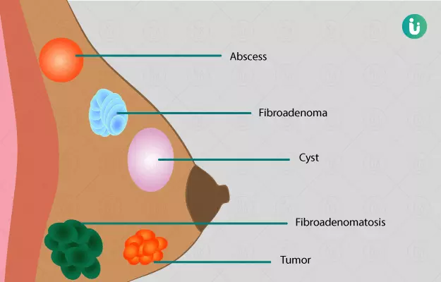
Breast lumps are abnormal tissue growths in the breasts. These may be round or irregular, painless or painful, with or without ulcers, soft or hard, and cancerous or non-cancerous. Many breast lumps are not harmful. Hence, when you identify or are diagnosed with a breast lump, do not worry. But make sure you visit a doctor to get the correct diagnosis.

Ignoring a breast lump is not a good idea because it may spread and cause discomfort. If left untreated, they may cause severe complications and you may have to get your breast removed.

  1. Non cancerous breast lumps
  2. Cancerous lumps
  3. How to check for breast lumps at home
  4. Breast lump symptoms
  5. Breast lump causes and risk factors
  6. Diagnosis for lump in breast
  7. Breast lump treatment

Non-cancerous breast lumps are those abnormal growths in the breasts that do not have cancer cells. They do not spread outside breast tissue and are not life-threatening.

  • Fibroadenoma
    fibroadenoma is the most common type of breast lump which occurs in women. This condition is characterised by an abnormal growth of both fibrous and glandular tissue of the breast. These lumps are usually soft to firm and movable in the breast. They are not fixed to the surrounding breast tissue.
  • Cysts
    Cysts are soft, fluid-filled sac-like growths, which are mostly round in structure. They may also cause slight pain in the breasts.
  • Fibrocystic disease
    Fibrocystic disease of the breasts involves three types of tissue damage which includes cyst formation, fibrosis (abnormal growth of fibrous tissue), and overgrowth of the tissues of breast glands.
  • Abscesses
    Abscesses occur because of an infection in the breast. They may sometimes be associated with ulceration in the breast skin. They are painful and discomforting. Abscesses most commonly occur in breastfeeding women.
  • Adenoma
    Adenomas are tumours, which occur when the inner lining or epithelium of the glands in the breast start growing abnormally.
  • Papilloma
    Papillomas are small finger-like projections that grow both inside and outside the milk ducts. They may also be associated with a discharge from the nipples. This discharge may also show signs of blood.
  • Lipoma and fat necrosis
    Lipoma refers to abnormal growth of fatty tissue inside the breast. Fat necrosis occurs when there is death and dissolution of the fat cells of the breast.
Women Health Supplements
₹719  ₹799  10% OFF
BUY NOW

Cancerous lumps are initially not painful but they grow rapidly and do not have well-defined boundaries. These growths may extend and destroy the adjacent tissues. They are usually hard and fixed to the surrounding or underlying breast tissue. If left untreated, some of the cancer cells may break off and travel to different parts of the body and cause cancer there. This is known as metastasis of cancer.

Read more: Symptoms of breast cancer

Breast self-examination is a good practice to know how your breasts feel. Hence, doing this every month will let you know if there is any abnormality in your breasts. Do your breast examination every month three to five days after your periods. You may follow these steps to examine your breasts yourself:

Lying down

Lying down helps in a better examination of your breasts and it’s easy to figure out if there is a lump in them. Lie down and put your left hand behind your head. With your right hand, start examining your left breast. In order to do so, use small, gentle yet firm, circular motions to check your breast. Start from the bottom of your breast and move upwards. Repeat the same procedure to examine your right breast and put your right hand behind your head.

Sitting

After completing the examination on lying down, sit up and start examining your armpits because your breast tissue also extends to each armpit. Use the same circular motions to feel the deeper tissues in your armpits for any abnormal growth or lump. You may also gently squeeze your nipple to see if there is any discharge from them.

Standing

You may do the examination in two ways; one with hands resting on your waist and another by raising them above your head. While you do that, look at your breasts in the mirror and check for the following things:

  • The outline of both your breasts should be more or less the same.
  • Check if the skin of both your breasts is similar and normal like the rest of the skin of your body.
  • Look for the presence of any dimpling or orange peel-like appearance in any of the breasts. If it’s there, you should consult a doctor.
  • The size of both your breasts should be similar.
  • The position of both your nipples should be at almost the same level.
  • See if there is any inward dipping or contraction in your nipples.
myUpchar doctors after many years of research have created myUpchar Ayurveda Prajnas Fertility Booster by using 100% original and pure herbs of Ayurveda. This ayurvedic medicine has been recommended by our doctors for lakhs of male and female infertility problems with good results.
Fertility Booster
₹899  ₹999  10% OFF
BUY NOW

You may experience one or more of the following symptoms of breast lumps:

  • Sudden enlargement of one of the breast, as compared to the other. 
  • Orange peel-like appearance of the skin around the nipple, and breast region. 
  • An abnormal shift in the position of one or both the nipples.
  • Discharge from nipples, which can be watery, whitish-yellow, greenish, brownish and reddish. Often reddish discharges are bloody in nature and should be reported immediately to a doctor. 
  • A dimple or depression of breast tissue in one or both the breasts.
  • You may also experience pain or heaviness in one or both the breasts.
  • Weight loss or appetite loss.
  • Appearance or feeling of a round mass or irregular soft to firm lump in your breast.

Causes

The causes of developing a breast lump include the following:

  • Infection
    Infection of the breast tissue either through the nipples or from an infection that is already in the blood circulation can cause the formation of a lump. An infection of the armpit may also cause infection in your breasts.
  • Inflammation
    Swelling of the breast tissue may cause pain or discomfort.
  • Trauma
    A sudden blow or injury to the breasts may cause the development of a mass, infection or swelling in the breast tissue.
  • Radiation
    If you are getting radiotherapy for a disease, your breast cells may have an abnormal overgrowth after getting damaged by the radiation.
  • Cancer-causing viruses
    There are some viruses and bacteria which alter the breast cells and result in their abnormal growth. These include human papillomavirus (HPV), Epstein-Barr virus, bovine leukaemia virus, etc.

Risk factors
There are certain factors that put you at a higher risk of developing breast lumps and breast cancer. These are:

  • Gender
    According to “An International Comparison of Male and Female Breast Cancer Incidence Rates”, women are more prone to developing breast lumps as compared to men owing to the greater level of estrogen in their body.
  • Obesity
    Being overweight not only increases your risk of developing a breast lump but is also related to a higher risk of getting heart diseases.

Read more: Obesity complications

  • Age
    A study, “Benign Breast Disease Heterogeneity: Association with histopathology, age, and ethnicity” suggests that elderly women above 56 years of age are at a lower risk of developing cancerous and non-cancerous lumps as compared to younger women. This is because the influence of sex hormones on the breasts is lowered after menopause (cessation of menstrual cycles). Also, the breast tissue starts regressing after this age and has a very low rate of cell growth. 
  • A family history of breast lumps or cancer
    If any member in your paternal or maternal or immediate family has had breast lumps or cancer, you are also prone to developing such diseases. These lumps are found to have a hereditary course as well.
  • Previous breast biopsies
    If you’ve had a previous biopsy of your breast tissue, it is possible that you might get a breast lump in association with that biopsy scar tissue.
  • Hormone replacement therapy
    A review of “Benign Breast Disease in Women” suggests that women who receive hormone replacement therapy after menopause are at a higher risk of developing breast lumps, especially those who start it within a short period of time after menopause.
  • Smoking and alcohol
    According to the “Recent insights into cigarette smoking as a lifestyle risk factor for breast cancer”, smoking and alcohol are amongst the factors which increase the risk of breast diseases and cancer in women who consume them in more than moderate amounts.
  • Gene mutation
    The genes BRCA1 or BRCA2 are associated with producing such proteins which help in suppressing tumours in the breast and ovaries. They also help in repairing the DNA and hence maintain the stability of the genetic material. Any mutation in these genes increases the risk of breast and ovarian cancer in women. 

    Find women health information

Diagnosis of breast lumps includes three stages commonly known as the “Triple Assessment”. These stages are clinical assessment, imaging, and biopsy.

Clinical assessment
This is the first stage of diagnosis in which a qualified doctor will ask you about the duration and symptoms associated with breast lumps. Your physician may also ask you about other things such as the age of onset of menstrual cycles, the age of menopause, number of children you have, any member in your family with breast cancer or any other cancer, etc. After that, your doctor will perform a detailed physical examination of your breasts to know the nature, size, and extent of the lump. If your doctor is a male, a female attendant or a nurse will be there during the examination.
Imaging
After the clinical examination, your physician may advise you to undergo one or a few of the following investigating procedures:

  • Mammography
    This is a procedure in which low-dose x-rays are used to get a picture of internal and external breast tissue. It is very helpful in identifying hidden cancerous growths and lumps. This procedure is also used as a screening method in which women above 30 years of age are investigated for the presence of unknown and asymptomatic breast lumps.
  • Ultrasound
    Ultrasound is usually done in women who are less than 30 years of age. In this procedure, sound waves are passed through your breast tissues which bounce back from the tissue to a probe. The latter then sends signals to an attached computer which forms images on a screen.
  • MRI (Magnetic Resonance Imaging)
    MRI is very sensitive in identifying breast cancer. It can detect both invasive and non-invasive types of breast lumps.

Biopsy
This is the third stage and is very specific and effective in diagnosing the type of breast lump. In this, a tissue sample from your breast is taken out and investigated to know the type of abnormality in your breasts. A biopsy can be done using a fine needle, core needle or excision.

  • Fine needle aspiration cytology (FNAC)
    In an FNAC test, a fine needle is used to withdraw cells and fluid from the affected area of your breast which is then are examined under a microscope.
  • Core needle biopsy
    This procedure is similar to that of fine needle biopsy. The only difference is that the needle used is slightly larger than that used in FNAC.
  • Excisional Biopsy
    In this procedure, a small amount of the affected tissue is cut from your breast using a germ-free blade. The tissue is then sent for processing in a lab where it is examined by an expert to diagnose the type of lump that you have in your breast.

Imaging for Metastases
These tests are done if the spread of cancer to other parts of the body or its metastases is suspected. A chest x-ray is done to determine the metastases in the lungs. CT scan of liver and abdomen may be helpful if metastases to these areas is suspected.

Ashokarishta
₹360  ₹400  10% OFF
BUY NOW

Medicines

Depending on the cause of your breast lump, your physician may prescribe you medicines to treat your condition.

  • Antibiotics and anti-inflammatory medicines
    Your physician may prescribe these medicines if the underlying cause of your breast lump is an infection or inflammation (swelling) in your breasts.
  • Chemotherapy
    Chemotherapy is a wide term that means treating a disease or disorder using medicines. However, this term is commonly used to indicate a treatment of tumours using medicines. Hence, if you have tumours in your breasts, these medicines are used to either dissolve them or reduce their size before surgery.

Radiotherapy

Radiotherapy uses radiation to kill the tumour cells to either resolve the lump or reduce its size before surgery.

Surgery

If the above methods do not regress your breast lump, or when cancer is diagnosed, you may have to undergo a surgery to get the tumour removed from your breasts. In severe cases, complete breast removal may be advised.

  • Lumpectomy
    In a lumpectomy, the surgeon removes the lump in your breast to prevent it from further growing and damaging the rest of the breast tissue.
  • Mastectomy
    Mastectomy is a procedure in which your breast is partially or completely removed when no other method is helpful in treating your condition. Sometimes, the lump may also spread to your armpits or underlying muscles. In that case, some part of these tissues is also removed along with the breast.

Read more: Breast cancer surgery

References

  1. Toomey A, Le JK. Abscess, Breast. [Updated 2019 Jan 11]. In: StatPearls [Internet]. Treasure Island (FL): StatPearls Publishing; 2019 Jan-.
  2. MedlinePlus Medical Encyclopedia: US National Library of Medicine; Breast self-exam
  3. Colin B Seymour, Carmel Mothersill. Breast cancer causes and treatment: where are we going wrong? Breast Cancer (Dove Med Press). 2013; 5: 111–119. PMID: 24648764
  4. Diana Ly, David Forman, Jacques Ferlay, Louise A. Brinton, Michael B. Cook. An International Comparison of Male and Female Breast Cancer Incidence Rates. Int J Cancer. 2013 Apr 15; 132(8): 1918–1926. PMID: 22987302
  5. Shannon Kispert, Jane McHowat. Recent insights into cigarette smoking as a lifestyle risk factor for breast cancer. Breast Cancer (Dove Med Press). 2017; 9: 127–132. PMID: 28331363
  6. Jingfang Cheng, Shijing Qiu, Usha Raju, Sandra R. Wolman, Maria J. Worsham. Benign Breast Disease Heterogeneity: Association with histopathology, age, and ethnicity. Breast Cancer Res Treat. 2008 Sep; 111(2): 289–296. PMID: 17917807
  7. Shannon Kispert, Jane McHowat. Recent insights into cigarette smoking as a lifestyle risk factor for breast cancer. Breast Cancer (Dove Med Press). 2017; 9: 127–132. PMID: 28331363
  8. Santen RJ. Benign Breast Disease in Women. [Updated 2018 May 25]. In: Feingold KR, Anawalt B, Boyce A, et al., editors. Endotext [Internet]. South Dartmouth (MA): MDText.com, Inc.; 2000-.
  9. National Cancer Institute [Internet]. Bethesda (MD): U.S. Department of Health and Human Services; BRCA Mutations: Cancer Risk and Genetic Testing
Read on app