Reproduction and conception of progeny is a natural process in the life cycles of all living beings. Humans are no different and pregnancies and childbearing is a life milestone many aspire to and desire. In order to understand how pregnancies occur and babies develop, it is paramount to understand the concept of fertilisation. Fertilisation in humans can simply be described as the fusion of the male gamete of sex cell called sperm with the female gamete or sex cell called the egg or ova.

(Read more: Pregnancy test kit)

Fusion of the male gamete cell (or sperm) nucleus with the female gamete cell (or ovum) nucleus results in fertilisation that gives rise to the zygote. After semen is deposited into the female genitalia (vagina or cervix), sperm enters the female reproductive tract and can survive in it for up to three days. The sperm swims through the cervix, into the uterus and then up into the fallopian tubes where the released ovum is usually located. On encountering the ova cell, the sperm releases chemicals that help it be identified by the ova. One ova can be fertilised by only one sperm even though it is likely to come in contact with many. After identification, the sperm releases chemicals that help it break the ovum cell membrane and enter it, allowing the nuclei of both cells to merge into one, thereby fertilising the ovum. The fertilised ovum is called the zygote. The zygote remains in the fallopian tube for about 72 hours and during this time it develops rapidly. If the fertilised zygote splits, it results in identical twins as the genetic material shared by the two newly formed zygotes is the same. Although usually only one ovum is released in each menstrual cycle by one of the ovaries, sometimes both ovaries may release an egg each. If two eggs are released in a cycle, they may both become fertilised by two different sperm cells, resulting in fraternal (unidentical) twins.

  1. Role of ovulation in fertilisation
  2. Role of sperm development in fertilisation
  3. What happens after fertilisation
  4. Applications associated with fertilisation
Doctors for Fertilisation

Ovulation is a crucial step in the normal female menstrual cycle, essential for the release of the fully mature egg into the female reproductive tract. The entire menstrual cycle is conducted by the interplay of hormones released from the hypothalamus, pituitary gland and the ovaries (also called the hypothalamus-pituitary-ovarian axis), which brings about changes in the uterus to be able to host a pregnancy, if conceived, and the ovaries to produce ova. The menstrual cycle can be described into two distinct phases, separated by the event of ovulation – the follicular phase and the luteal phase. Changes occur in the innermost uterine layer, called the endometrium, at the same time as the changes in the ovaries.

  • Follicular phase: Immediately after a menstrual period of bleeding ends, the next menstrual cycle begins. The first phase of the menstrual cycle is known as the follicular (or proliferative) phase. The hypothalamus regulates the release of pituitary hormones. The pituitary is the master gland of the body that regulates the release of all other hormones in the body in a timely manner. During the first follicular phase, the hypothalamus signals the pituitary to release two hormones – the Follicular Stimulating Hormone (FSH) and Luteinizing Hormone (LH). These two hormones stimulate the ovarian follicles that contain the developing egg cells within them; hence, this phase is called the follicular phase. The concentrations of FSH and LH remain high and steady throughout the follicular phase. The growing ovarian follicles produce large amounts of estrogen, but no progesterone is produced in the ovaries at this time.
  • Ovulation: As the ovarian follicles develop completely, the level of estrogen being produced by them rises. High circulating estrogen levels send a signal to the pituitary to secrete even more FSH and LH. This LH surge is responsible for the rupture of the fully developed ovarian follicle, thereby releasing the fully developed ova from within it into the female reproductive tract. Following ovulation, the ovum is picked up by the fimbriae of the fallopian tube on the same side of the body as the ovary that released the ovum. Depending on the length of the menstrual cycle, ovulation varies. The ovulation event usually occurs 14 days before the start of the next menstrual period in menstrual cycles of an average length of 21 to 35 days. After the release of the ovum, it can survive in the female reproductive tract for around 24 hours; this is known as the “fertility window” when unprotected sexual intercourse has the highest chance of resulting in a pregnancy.
  • Luteal phase: After the ovarian follicle has ruptured and the ova has been released, the estrogen levels fall sharply as the follicles that produced it are no more. This signals to the pituitary gland to not release any more FSH or LH, as they are no longer needed. The cells of the ruptured follicle reorganise themselves into a special structure called the corpus luteum, thus giving the second phase of the menstrual cycle the name of the luteal phase. The role of the corpus luteum is to produce hormones, progesterone and small amounts of estrogen, in order to maintain the uterine endometrial integrity to sustain a pregnancy that may be conceived. Two events can occur:
  • If a pregnancy is not conceived, the corpus luteum in the ovary regresses, resulting in levels of progesterone and oestrogen dropping. As these two hormones are necessary to keep the endometrium lining of the uterus intact, the endometrium breaks down and is expelled as menstrual blood.
  • If pregnancy is conceived, the corpus luteum develops further into a structure called the corpus luteum of pregnancy and produces even larger amounts of progesterone and estrogen to sustain the uterus and pregnancy.
Women Health Supplements
₹719  ₹799  10% OFF
BUY NOW

While the ovum is being prepared in the female reproductive tract, sperm (the male gametes) is being prepared in the male reproductive tract. After males attain puberty, sperm begins to develop in the male gonads called testes. The two testes are located inside two separate pouch-like structures called the scrotum, which is located outside the pelvic cavity, below the male genitalia or penis. Inside each testis is a network of tubelike structures called the seminiferous tubules. It is in these seminiferous tubules that the sperms are produced from immature cells called spermatogonia. Much like the female gamete cycle, spermatogenesis, or spermatozoa production in males is controlled by the hypothalamus, pituitary and testes. The hypothalamus signals to the pituitary gland to release FSH and LH at the appropriate time in males. FSH acts on the Sertoli cells of the testes and LH acts on the Leydig cells to produce testosterone. Immature sperm cells develop into spermatozoa, or sperms, and have a flagellum or tail that helps them move. Testosterone and inhibin are two hormones that are formed in the testes under the influence of FSH and LH, which signal to the pituitary and the hypothalamus to stop further hormone release after the sperm is ready. The flagellated (possessing tails for movement) sperms travel from the testes into the epididymis. When the male is stimulated sexually, sperm is released along with secretions of other glands (like seminal vesicles, prostate and Cowper’s glands) as a white fluid called semen. After semen is deposited in the female genitalia (through the vagina or cervix), following unprotected sexual intercourse or artificial insemination, sperm enters the female reproductive tract. Sperm can survive in the female reproductive tract for up to 3 days. If it encounters the egg cell within this duration, chances of fertilisation are high.

(Read more: Semen analysis)

Once the sperm fertilises the ova, other changes begin to take place. Following is how the development begins:

  • Embryo: Until the eighth week of pregnancy, the developing human is called an embryo. After fertilisation, the zygote grows and differentiates rapidly. First, it divides into two cells; then they each divide again to give four cells in total. The process of cell division is repeated many times until a solid ball of cells called a morula is produced after about three days. As the cells in the morula continue to divide, they begin to move towards the outer edges of the ball formed in the morula stage; this results in the “ball” becoming a hollow ball of cells called the blastocyst at about five days after fertilisation.
  • Implantation: Between five to seven days after fertilisation, the blastocyst formed from the fertilised zygote reaches the uterus from the fallopian tube and embeds itself in the thickened endometrial lining of the uterus. This process is called implantation and, if the embryo survives it, is the beginning of a pregnancy. If the blastocyst has implanted successfully into the uterus, it will grow further.  Three or four blastocyst cells develop into the inner cell mass, which will eventually give rise to the fetus or baby, and the remaining 100 or so blastocyst cells form a structure called the trophoblast, which will provide the baby’s contribution to the placenta.
  • Sometimes, the blastocyst may implant into tissue outside the uterus (like the Fallopian tubes) resulting in an ectopic pregnancy. Ectopic pregnancies do not reach term or result in the delivery of a baby. Ectopic pregnancies are usually removed surgically. Sometimes, growing ectopic pregnancies can rupture and result in internal bleeding and hypovolemic shock.
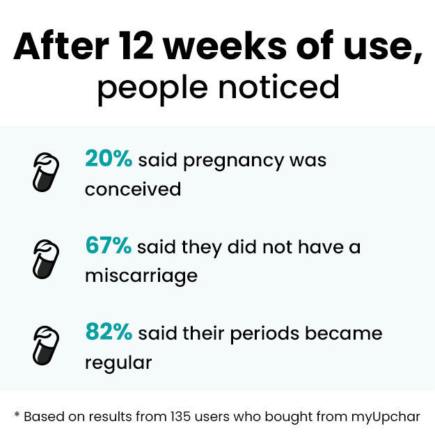
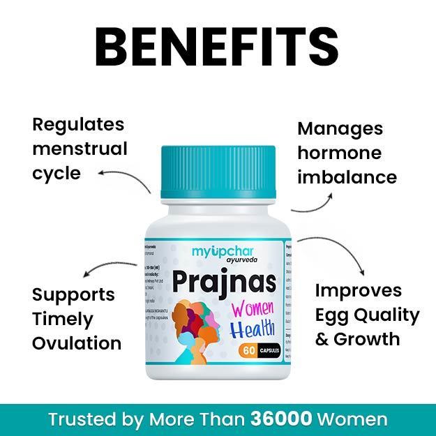
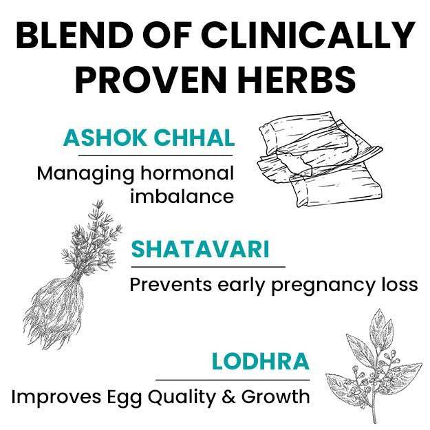
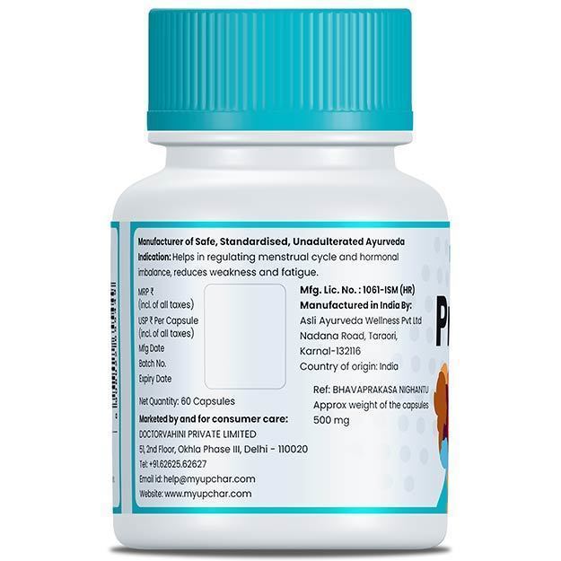
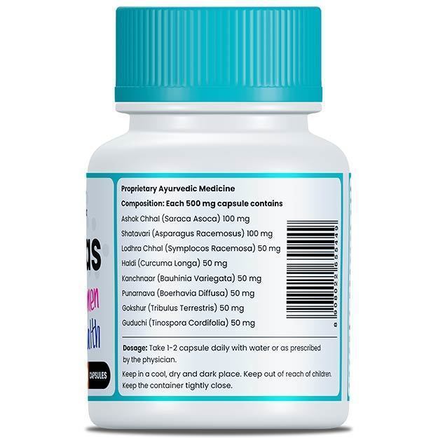
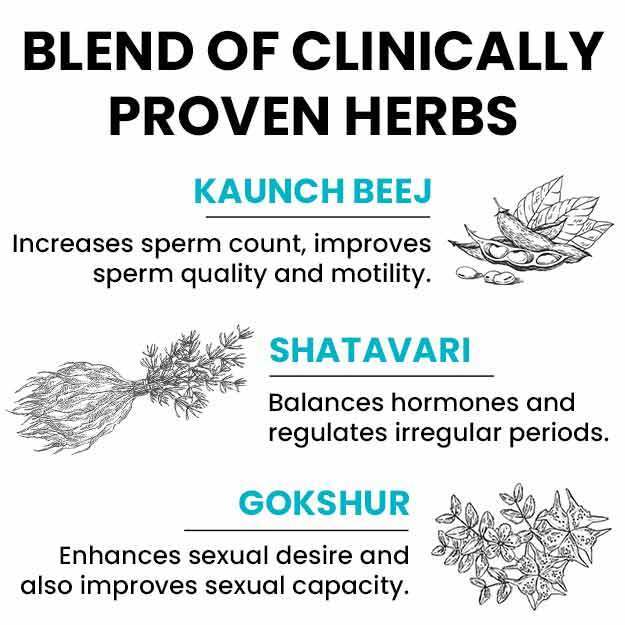
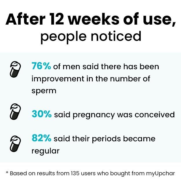
myUpchar doctors after many years of research have created myUpchar Ayurveda Prajnas Fertility Booster by using 100% original and pure herbs of Ayurveda. This ayurvedic medicine has been recommended by our doctors for lakhs of male and female infertility problems with good results.
Fertility Booster
₹899  ₹999  10% OFF
BUY NOW

After understanding how fertilisation occurs, we can discuss its implications in medicine and daily life.

  • Contraception or preventing fertilisation: In order to prevent conceiving unwanted pregnancies, any step of the process leading up to fertilisation can be inhibited. For example, the following strategies are usually employed for contraception:
    • Ovulation inhibition: Oral contraceptive pills inhibit ova formation. 
    • Vasectomy: Older males who do not wish to bear any more children can undergo a procedure to prevent sperm release in semen. 
    • Barrier methods: Condoms, for instance, are the most commonly used contraceptive method, that prevents sperms and semen from entering the female reproductive tract to prevent fertilisation.
  • Infertility: Primary infertility is defined as the failure to conceive a child after regular unprotected sexual intercourse over a period of 1 year (if the female partner is less than 35 years of age) or 6 months (if the female partner is more than 35 years of age), having had no previous pregnancies, irrespective of the outcome. The failure for fertilisation to occur is the basis of infertility. Reasons for infertility can therefore be due to any factor in the fertilisation process. Such as:
    • Ovulation problems 
    • Sperm problems 
    • Sexual problems
  • In Vitro Fertilisation (IVF): Some patients who are unable to conceive naturally, require assisted reproductive technology (ART). If fertilisation is unable to occur in the female body after regular unprotected sexual intercourse over a period of time or after artificial insemination with donor sperms, in vitro fertilisation (IVF) is attempted. Eggs are retrieved from the female donor and mixed with sperm from a male donor in a Petri dish in a laboratory to achieve fertilisation. If fertilisation occurs successfully, the zygote is allowed to grow suitably and the embryo is implanted into the female who will carry the pregnancy to term.
Dr. Arpan Kundu

Dr. Arpan Kundu

Obstetrics & Gynaecology
7 Years of Experience

Dr Sujata Sinha

Dr Sujata Sinha

Obstetrics & Gynaecology
30 Years of Experience

Dr. Pratik Shikare

Dr. Pratik Shikare

Obstetrics & Gynaecology
5 Years of Experience

Dr. Payal Bajaj

Dr. Payal Bajaj

Obstetrics & Gynaecology
20 Years of Experience

Read on app