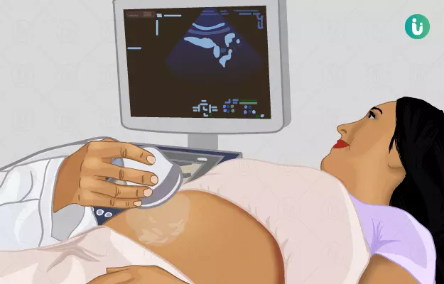
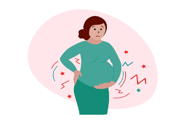
Getting an ultrasound done is indeed an emotional time for the parents as this allows them to see their baby even before they can hold it in their arms. A pregnancy ultrasound, also known as a sonogram is a prenatal test advised to most expecting mothers to check the baby's health and growth.

The test uses high-frequency sound waves to show images of your baby, placenta and ovaries during varied times of your pregnancy. The sound travels through a probe that is passed over the belly of the mother and comes back to the probe after bouncing back from body tissues. An attached computer records the echoes and transforms them into pictures or videos.

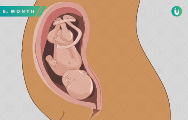
You may be able to see your baby’s hands, legs and other body parts, hear the baby’s heartbeat, and see him swim inside your womb depending upon when the ultrasound has been done. Ultrasound can also tell you if it is a boy or a girl. Although in India and in many other countries, the revelation of sex is unethical and prohibited.

  1. Why is pregnancy ultrasound or pregnancy sonography done?
  2. Types of ultrasound during pregnancy
  3. How is pregnancy ultrasound done?
  4. How many ultrasounds are done during pregnancy?
  5. Ultrasound scan in case of twins or triplets
  6. How to prepare for the pregnancy ultrasound?
  7. Are ultrasounds during pregnancy safe?
  8. Takeaway on ultrasound during pregnancy

Ultrasound during pregnancy is done multiple times during pregnancy. The first is to confirm the pregnancy and to find out the date of pregnancy. Further, the procedure is carried out to see the growth and development of the foetus, and to check the progress of pregnancy. During a certain point in the second trimester, ultrasound helps in looking at the anatomy and heartbeat of your baby.

Various reasons why pregnancy ultrasound is done can be compiled as:

  • Ultrasound as a pregnancy test: Ultrasound is done to confirm a suspected pregnancy. During this time, it also helps to determine the presence of an ectopic pregnancy (fetus developing in fallopian tubes). (Read more: pregnancy test)
  • Ultrasound to check baby’s age during gestation: Gestational age of baby can be determined by identifying the gestational sac in the first trimester. Transvaginal ultrasound can determine the pregnancy age as early as 4 weeks and transabdominal ultrasound can identify the pregnancy age as early as 5 weeks. This helps in estimating the due date of delivery.
  • Ultrasound to confirm the number of pregnancies: Ultrasound also helps to confirm the number of babies, i,e if you carrying a single child, twins or triplets. This is usually revealed when you are 10 weeks to 13 weeks pregnant.
  • Ultrasound to assess the fetal growth: Ultrasound helps to assess the growth of the baby during the second and third trimesters of pregnancy. The scan helps to see the arms, legs and other body parts of a baby and helps the doctor to evaluate if baby measures bigger or smaller than expected gestational age.
  • Ultrasound to identify birth anomalies: Mid-pregnancy ultrasound helps your doctor to rule out if there are any birth anomalies such as cleft lip (openings or divisions in the mouth and lip roof), heart and brain anomalies or spina bifida (baby’s spinal cord fails to develop properly).
  • Ultrasound to check the amniotic fluid and placenta: Baby is connected to placenta via the umbilical cord and obtains all vital nutrients through it. Ultrasound helps to determine the placenta position and examines if it is positioned close or away from the cervix. An ultrasound also helps to assess the amniotic fluid levels in the body. Amniotic fluid surrounds the fetus and protects the baby against any pressure and keeps the temperature consistent.
  • Examining baby’s position inside the womb: Ultrasound helps to find out the position of your baby. Your doctor can assess if your baby is head up and bottom down or baby’s legs are stretched out or bent. This helps to decide if the delivery of the baby would be normal or a caesarean. (Read more: Normal delivery)
Women Health Supplements
₹719  ₹799  10% OFF
BUY NOW

The following are some types of ultrasounds that can be done during pregnancy

  • Transvaginal ultrasound during pregnancy: During pregnancy, when the baby is too small, a transvaginal ultrasound or endovaginal ultrasound is recommended to get a better picture of the baby. The procedure involves the insertion of an ultrasound probe 2 to 3 inches inside the vagina, while the transducer is kept outside of the pelvis. This is usually done to examine if there is any vaginal bleeding, monitoring the heartbeat of the baby, examining the placenta, confirming early pregnancy or doubtful miscarriage. (Read more: Early signs of pregnancy)
  • Standard transabdominal ultrasound during pregnancy: Transabdominal ultrasound is a standard ultrasound that evaluates the fetus at 8 weeks gestation. In this procedure, a transducer connected to a monitor is passed over the abdomen of the woman. The transducer produces sound waves, that echo back and form the image on the monitor.
  • Doppler ultrasound during pregnancy: Doppler ultrasound utilises sound waves or echoes to examine the blood flow in vessels. Doppler ultrasound during pregnancy helps to assess blood circulation in the baby, uterus and placenta.  
  • 3-D ultrasound during pregnancy: A 3D ultrasound is a transabdominal ultrasound that takes 2-D pictures of the baby which are then translated by computer into 3-dimensional images that come as a real-life photograph. Although the 3-D images look much fascinating, studies show mixed results on the safety of taking a 3-D ultrasound.
  • 4-D ultrasound during pregnancy: Unlike 2-d or 3-D images, 4-D ultrasound uses the scanners that show up the movement of the baby in addition to the images.
  • Fetal echocardiography during pregnancy: Using this technique, doctors can assess the heartbeat of a baby. This test is mostly performed to check congenital heart diseases. (Read more: Fetal echo procedure)

In case of conventional ultrasound, your doctor would probably ask you to first change into a gown and lie on a padded examination table. A small amount of water-soluble gel will be applied to your abdomen. Then a small device called a transducer is passed over the abdomen. The transducer sends sound waves into your tummy, reflecting out your baby’s images.

In general, there is no discomfort felt during the procedure. The gel is wiped off using a towel or tissue after completing the procedure.

This is the question that every expecting woman is bothered about. No recommended number of ultrasounds have been recorded for routine prenatal care. And because ultrasounds do not have any scientifically proven issues and are relatively safe, these can be carried out at any time during pregnancy.

Usually, ultrasound is done every trimester to evaluate baby’s health anD mother’s womb.

  • Ultrasound during the first trimester of pregnancy: During the first trimester of pregnancy, ultrasound is done to check the viability of the fetus, find the due date, examine the baby’s heartbeat, check the number of pregnancies, positioning, and to rule out any vaginal bleeding. This is usually done when you’re 6-8 weeks pregnant.
  • Ultrasound during the second trimester of pregnancy: During the second trimester of pregnancy, an anatomy ultrasound is done to examine the baby’s growth, to assess any birth defects or congenital diseases, baby’s anatomy and baby’s gender. Determination of sex is prohibited in India. A second ultrasound is done at 18-20 weeks of pregnancy.
  • Ultrasound during the third semester of pregnancy: Ultrasound in the third trimester of pregnancy is done to evaluate the baby’s growth, any anomalies, or any changes in baby’s positioning. This is done at 32-34 weeks of pregnancy.

(Read more: Checkups during pregnancy)

myUpchar doctors after many years of research have created myUpchar Ayurveda Prajnas Fertility Booster by using 100% original and pure herbs of Ayurveda. This ayurvedic medicine has been recommended by our doctors for lakhs of male and female infertility problems with good results.
Fertility Booster
₹899  ₹999  10% OFF
BUY NOW

According to the National Institute for Health and Care Excellence, women who are pregnant with twins or triplets (known as multiple pregnancies) are recommended to get an ultrasound scan done between 11 weeks and 13 weeks 6 days of their pregnancy to record the positions of their babies. Also, the doctors make sure that they label the foetuses in case of multiple pregnancies, using an ultrasound scan. Women with multiple pregnancies are recommended to have details scans every 4 weeks after the second trimester to examine any structural abnormalities.  

(Read more: Twin pregnancy symptoms)

It is easy to prepare for an ultrasound. You need to get your bladder full in order to get clear scans and high-quality pictures of the baby. So make sure you drink 8 glasses of water or more before you go for sonography. Just that your bladder should be full and you do not have to pass the urine, no matter how urgent you feel.

Ultrasound is the most common procedure in pregnancy diagnostics. Undoubtedly it is a convenient and painless procedure, but women are still bothered about the safety of these scans. According to the American Pregnancy Association, “The long-term effects of repeated ultrasound exposures on the fetus are not fully known. It is recommended that ultrasound be only used if medically indicated.”

Ashokarishta
₹360  ₹400  10% OFF
BUY NOW

A pregnancy ultrasound is a prenatal test that involves the use of high-frequency sound waves to get the images of a baby and mother’s womb. Surely, this is an exciting time for the parents but go for an ultrasound when medically recommended. Even if the scans are considered safe, you should still use ultrasound sparingly, and avoid needless scans. Go for an experienced ultrasound specialist, and opt for procedures that give good results while giving less exposure.

References

  1. National Institute for Health and Care Excellence. Multiple pregnancy: twin and triplet pregnancies. [Internet]
  2. A. Khalil et al. ISUOG Practice Guidelines: role of ultrasound in twin pregnancy. Ultrasound in Obstetrics & Gynecology
  3. American Pregnancy Association. [Internet]; Ultrasound: Sonogram.
  4. Better health channel. Department of Health and Human Services [internet]. State government of Victoria; Pregnancy tests - ultrasound
  5. U. S Food and Drug Association. [Internet]. Ultrasound Imaging
  6. MedlinePlus Medical Encyclopedia: US National Library of Medicine; Ultrasound pregnancy
  7. Office on Women's Health [Internet] U.S. Department of Health and Human Services; Prenatal care and tests.
Read on app