Pericardial effusion

Dr. Suvansh Raj NirulaMBBS

November 20, 2020

January 08, 2021

Pericardial effusion

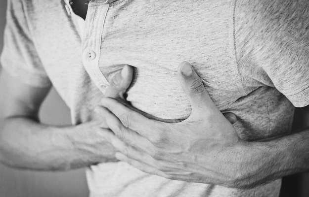
An abnormal accumulation of fluid in the pericardial space in the chest cavity is known as pericardial effusion.

The pericardium is a fluid-filled sac-like structure that envelops the heart and the roots of major vessels. These major vessels are the aorta, the pulmonary artery, the pulmonary veins, the superior vena cava and the inferior vena cava.

The pericardium is composed of two major layers. They are: 

  • Fibrous pericardium: It is basically a continuation of the tendon of the diaphragm. It is formed by connective tissue. This tough tissue prevents the overfilling and the overexpansion of the heart. 
  • Serous pericardium: This layer itself is composed of two layers. 
    • Outer parietal layer: Lines the inner side of the fibrous pericardium
    • Internal visceral layer: Lines the outer surface of the heart. This layer is also called the epicardium.

Both these layers are formed by a single layer of epithelial cells (cells that line the surfaces of the body and act as a shield to protect the inside of the body from the germs present in the environment). This single layer is termed as the mesothelium.

Pericardial cavity/pericardial space is present between the outer parietal layer and the inner visceral layer of the serous pericardium. This space consists of lubricating fluid that helps in reducing the friction produced by the contraction of the heart. 

Read on to know what causes excess buildup of fluid in this cavity or pericardial effusion, symptoms of pericardial effusion, diagnosis of pericardial effusion and treatment of pericardial effusion.

Is pericardial effusion common?

Studies suggest that small asymptomatic pericardial effusions have been discovered in nearly 3-3.5% of general autopsies. 

In the case of HIV (Human immunodeficiency virus) patients, before the highly active antiretroviral therapy (HAART) was introduced, nearly 11% of patients had some degree of pericardial effusion. After the discovery and prescription of HAART, this number has been brought down considerably. 

In patients suffering from any form of cancer, around 11% of autopsies suggested a pericardial effusion.

myUpchar doctors after many years of research have created myUpchar Ayurveda Urjas Capsule by using 100% original and pure herbs of Ayurveda. This ayurvedic medicine has been recommended by our doctors to lakhs of people for sex problems with good results.
Long Time Capsule
₹719  ₹799  10% OFF
BUY NOW

Symptoms of pericardial effusion

The symptoms of pericardial effusion may depend on the size and how fast the effusion happens. The following clinical features may be observed:

  • Small-sized effusions may be asymptomatic. They usually develop over a long period of time.
  • Chest pain: A sensation of pressure and discomfort in the chest. This pain increases on lying down and reduces on leaning forward
  • Fainting
  • Dizziness
  • Cough
  • Breathlessness
  • Confusion 
  • Anxiety 
  • Increased heart rate
  •  Generalised body swelling
  •  Increased size of the liver 
  •  Enlarged spleen
  •  Increased respiratory rate
  •  Pericardial friction rub: This is seen when the pericardial effusion is associated with inflammation of the pericardium. It is a high pitched scratching sound that increases on leaning forward while the patient breathes out. 
  •  Beck’s triad of cardiac tamponade: Cardiac tamponade may occur when the pericardial effusion compresses the heart muscles. The beck’s triad includes low blood pressure (hypotension), muffled heart sounds and distension of neck veins (jugular venous distension). 
  •  Poor peripheral pulses
  •  Pulsus paradoxus: Normally, systolic blood pressure or the upper number on a blood pressure machine reading drops up to 10 points during the inspiration phase (when you breathe in). A drop of more than 10 mm Hg or millimetres of mercury is known as a paradoxical pulse. This symptom helps in detecting the presence of cardiac tamponade in patients with pericardial effusion. 
  •  Bluish discolouration of skin and mucus membranes like the inside of the mouth. It occurs due to low blood oxygen levels. This condition is termed as cyanosis.

Pericardial effusion causes

There are numerous causes for the development of pericardial effusion. They are as follows: 

  • Idiopathic: Idiopathic diseases are those conditions that occur spontaneously and the reason for their occurrence is unknown.
  • Infections: An infection may lead to infectious pericarditis (inflammation of the pericardium). These infectious agents may be viral (as seen in human immunodeficiency virus/HIV infection), fungal, parasites or bacterial including syphilis and Mycobacterium tuberculosis (the causative agent for tuberculosis).
  • Acute myocardial infarction: Commonly referred to as a heart attack, it is a condition which involves the death of heart muscles due to limited blood supply.
  • Acute kidney failure
  • Chronic kidney disease
  • Malignancies (cancer): Pericardial effusion may occur due to metastatic spread of the primary cancer or opportunistic effusion that may occur due to a reduced immune response in cancer patients. Sometimes, pericardial effusions may develop due to a complication of cancer treatment such as radiotherapy or chemotherapy.
  • Hypothyroidism or low blood thyroid levels
  • Trauma
  • Severely low haemoglobin levels (severe anaemia)
  •  Sarcoidosis (a disease which involves the collection of inflammatory cells in a large number of lumps termed as granulomas)
  •  Rupture of aortic aneurysm (ballooning of a section of the aorta is termed as an aortic aneurysm)
  •  Autoimmune diseases: Ailments in which the body’s immune system attacks the body are known as autoimmune disorders. Examples include systemic lupus erythematosus (SLE)rheumatoid arthritis (RA), ankylosing spondylitis, scleroderma and acute rheumatic fever.
  •  Drug-induced pericardial effusion: Various drugs may lead to pericardial effusion such as hydralazine (for hypertension), isoniazid (for tuberculosis treatment), minoxidil (used in the treatment of male pattern baldness), anticoagulants and phenytoin (for seizures).
  •  An open heart surgery may lead to a pericardial effusion

Prevention of pericardial effusion

A pericardial effusion can be prevented if susceptible patients make drastic lifestyle changes, including:

  • Reduce alcohol intake
  • Consume a low-fat and low-salt diet
  • Get adequate exercise 
  • Lose weight
  • Treat underlying comorbidities
myUpchar doctors after many years of research have created myUpchar Ayurveda Kesh Art Hair Oil by using 100% original and pure herbs of Ayurveda. This Ayurvedic medicine has been recommended by our doctors to more than 1 lakh people for multiple hair problems (hair fall, gray hair, and dandruff) with good results.
Bhringraj Hair Oil
₹599  ₹850  29% OFF
BUY NOW

Pericardial effusion diagnosis

A diagnosis of pericardial effusion should be made based on the clinical presentation of the patient, biochemical and radiological investigations.

The biochemical investigations of a pericardial effusion should include the following: 

  • Increased white blood cell (WBC) count in a complete blood count would indicate an infection
  • Kidney function test and electrolyte levels can help a doctor determine whether blood urea levels are higher
  • Thyroid function test: The most important component of a thyroid function test is the thyroid-stimulating hormone (TSH) level.
  • Auto-antibodies: The presence of rheumatoid factor is suggestive of rheumatoid arthritis/RA or the presence of an anti-nuclear antibody may be suggestive of systemic lupus erythematosus (SLE) (read more: Rheumatoid arthritis factor test)
  • Cardiac markers: In case of certain complications like pericarditis, the tests might show elevated troponin levels.
  • Blood culture test, to detect the presence of any bacteria in the blood of the patient.
  • Carcinoembryonic antigen levels: Elevated levels may be seen in the case of malignant cases with pericardial effusion (read more: Carcinoembryonic antigen test)
  • Aspiration of pericardial fluid, to be sent for cytological and microbiological analysis. The level of proteins, white blood cells (WBCs) have to be assessed and cultures have to be performed.
  • A smear for the presence of acid-fast bacilli in patients suspected to suffer from tuberculosis and patients suffering from human immunodeficiency virus (HIV)  may also be done.

The radiological investigations that should be performed include the following: 

  • Electrocardiogram (ECG): An electrocardiogram (ECG) might show the presence of important ECG changes and signs that may be suggestive of a myocardial infarction or pericarditis. The main ECG change observed is the presence of raised ST segments.  Other changes include PR segment depression and bundle branch blocks.
  • Echocardiogram: A transoesophageal echocardiography may help in identifying a pericardial effusion and thickening. It may also be able to detect underlying metastatic causes. (For this test, the doctor will guide a long thin camera or endoscope through the throat to look at the heart).
  • Chest X-ray: Chest X-rays may show an increased heart size with sharp margins. This is termed as cardiomegaly. There may also be a presence of translucent lines in the cardiopericardial shadow. Chest X-rays are considered to be an important initial investigation as, apart from helping in the diagnosis of pericardial effusion, they also help in ruling out other dangerous and important causes of chest pain and breathlessness such as pneumothorax (collapse of the lungs). 
  • Magnetic resonance imaging (MRI) is considered to be a highly accurate and appropriate investigation to detect the presence of pericardial effusion. It can detect the presence of fluids (even in amounts as small as 30 ml). Magnetic resonance imaging utilizes magnetic rays to view, isolate and observe the problematic regions of the body.
  • Computed tomography (CT scan) is also considered to be very accurate and useful in cases of pericardial effusion. It is based on the generation of highly detailed body images using X-rays.
  • Pericardioscopy along with a pericardial biopsy: Pericardioscopy is a procedure in which the doctor can inspect the pericardium with the help of a camera. A pericardioscopy may also involve taking a pericardial biopsy to rule out malignancies in case a cancer is suspected. 

Differential diagnosis

Some conditions need to be considered and ruled out when making a diagnosis of pericardial effusion. They include the following: 

  • Cardiac tamponade: Compression of the heart due to a pericardial effusion
  • Pulmonary embolism: A blood clot in the pulmonary artery (major blood vessel that carries blood from the heart to the lungs). 
  • Myocardial infarction or heart attack
  • Cardiomyopathy is a disease that affects the heart muscles and makes it difficult for the heart to pump blood. This condition may lead to heart failure
  • Ventricular aneurysm: Ballooning of the weakened heart muscle. It is filled with blood. 

Pericardial effusion treatment and management

After the successful diagnosis of a pericardial effusion based on clinical features and investigations, cardiac tamponade must be ruled out. 

The treatment of pericardial effusion may be pharmacological or surgical or both. The treatment would depend on the size of the effusion, the speed of development and the patient profile along with the comorbidities (diseases that occur simultaneously and are often risk factors for each other) the patient might be suffering from. 

Patients with small-sized asymptomatic pericardial effusions who are clinically stable may be managed with a frequent follow-up clinical examination along with echocardiography.

Pharmacological treatment for pericardial effusion

From supportive therapies to medicines, the following medical interventions may be used in the treatment of pericardial effusion:

  • Oxygen supplementation: Helpful in clinically unstable patients with low blood oxygen levels (hypoxemia).
  • Treating the cause of pericardial effusion—for example, chemotherapy for patients suffering from any kind of cancer
  • Intravenous fluids to dehydrated patients with signs of low body fluid levels such as low blood pressure (hypotension).
  • Supportive treatment such as painkillers for chest pain.
myUpchar doctors after many years of research have created myUpchar Ayurveda Urjas Energy & Power Capsule by using 100% original and pure herbs of Ayurveda. This Ayurvedic medicine has been recommended by our doctors to lakhs of people for problems like physical and sexual weakness and fatigue, with good results.
Power Capsule For Men
₹719  ₹799  10% OFF
BUY NOW

Surgery for pericardial effusion

In patients who don’t respond well to supportive therapies, a surgeon can drain the extra fluid (pericardiocentesis). In cases where even that doesn’t work, there is the option of a pericardiotomy:

  • Pericardiocentesis is defined as the surgical drainage of fluid present in the pericardial space. It is not considered to be appropriate in patients affected by small pericardial effusions that may resolve spontaneously or with medical and supportive management.
  • Pericardiocentesis may not be performed in patients with ruptured ventricular aneurysms (ballooning of the weakened areas in the ventricles of the heart) or in the case of aortic dissection (tear in the innermost layer of the wall/tunica intima of the aorta)
  • Surgical approach (Pericardiotomy): Usually performed in patients with immense and chronic pericardial effusions. These patients often have a history of failed pericardiocentesis. 

Advantages of this method include low morbidity, low mortality along with a low recurrence rate. It also does not require general anaesthesia and can be performed using local anaesthesia.

Complications of pericardial effusion

In some cases, pericardial effusion may have complications like:

  • Cardiac tamponade: Compression of the heart muscles due to the development of pericardial effusion. It is an emergency condition that can sometimes lead to dangerous consequences including low blood pressure (hypotension), poor blood supply to vital organs and death. 
  • Chronic pericardial effusion: Pericardial effusion that lasts for a long time, usually beyond six months. This complication is not as dangerous as cardiac tamponade.

Prognosis for pericardial effusion

The prognosis of patients with pericardial effusion mainly depends on the reason behind it. 

Small size effusions may be tolerated well. They are usually caused due to non-life-threatening causes. 

Large pericardial effusions may be suggestive of more severe disease. They can cause serious complications including the death of the patient.

Follow-up after pericardial effusion treatment

The follow-up of pericardial effusion depends on the size of effusion measured on an echocardiogram along with levels of inflammatory markers such as erythrocyte sedimentation rate (ESR test) and C-reactive protein (CRP test). 

The monitoring regimens are as follows:

  • Small pericardial effusions (<10mm): Don’t require any monitoring
  • Moderate to large pericardial effusions (>10mm): Patient is to be followed up after one or two weeks. The next visits are in one month and six months. 

In case of severe effusions, an echocardiogram should be performed every three to six months.


Medicines for Pericardial effusion

Medicines listed below are available for Pericardial effusion. Please note that you should not take any medicines without doctor consultation. Taking any medicine without doctor's consultation can cause serious problems.