Download App


What is Fatigue Panel?
Fatigue is not a condition but a symptom. It is known as the feeling of being constantly tired. However, fatigue is more than just feeling sleepy and tired. If you find it difficult to carry on with your daily tasks and focus on the activities at hand even when you have slept enough, exercise regularly and take care of proper nutrition, you are experiencing fatigue.
Most adults experience it at some point in their life. Fatigue may be physical, mental or both. It may be either due to a medical condition or problems with the lifestyle or general and psychological wellbeing.
The tests in the fatigue panel include the following:
- Complete blood count (CBC): Doctors order this test when they notice symptoms such as fatigue, weakness and bruising. A CBC can help in diagnosing the conditions that cause these symptoms. It includes the following test:
- White blood cell (WBC) count: WBCs play a role in protecting your body against infection- both bacterial and viral and from other organisms. When you have an infection, the number of WBCs in your body rises. Thus, a WBC count helps in determining if you have an infection. In case of cancer patients, this test also helps determine how the body is responding to cancer treatment.
- Differential WBC count (DLC): There are various types of WBCs in the body - neutrophils, eosinophils, basophils, monocytes and lymphocytes. Different WBCs have different functions in protecting the body. This test checks for the number of each WBC in the body. Increase or reduction in any type of WBC can indicate specific conditions. For example high eosinophils are seen in allergies. A differential WBC count also helps determine the number of immature neutrophils called band neutrophils.
- Red blood cell (RBC) count: RBCs transport oxygen from the lungs to different regions of the body and carry carbon dioxide back to the lungs. If your RBC levels are too high (polycythaemia), there is a possibility of the RBCs to clump together and block the capillaries, which are tiny blood vessels. If your RBC levels are too low (anaemia), it could mean the body is not getting enough oxygen.
- Haematocrit (HCT): This test checks for the volume occupied by RBCs in blood. It is measured as a percentage of RBCs in a volume of blood. For example, if the HCT value is 42, it shows that the RBCs occupy 42% of the blood.
- Haemoglobin (Hgb): Hemoglobin is a protein present in RBCs. It is what carries and transports oxygen. Hgb is also responsible for imparting the distinct red colour to RBCs. The Hgb test checks for the body’s ability to carry oxygen. Along with HCT, this test can detect if you have anaemia and polycythaemia - both of which have fatigue as a symptom.
- RBC indices: RBC indices are values that are calculated using results of the other tests in CBC. There are three RBC indices: mean corpuscular volume (MCV, size of RBCs), mean corpuscular haemoglobin (MCH, amount of Hgb in an average RBC) and mean corpuscular haemoglobin concentration (MCHC, the concentration of Hgb in an average RBC). These indices can diagnose different types of anaemia.
- Red cell distribution width (RDW): This test checks if the size and shape of all the RBCs in your blood are the same or different.
- Platelet count: Platelets are the smallest blood cells that are essential for blood clotting. If your platelet count is low, you will have uncontrolled bleeding; whereas, if you have a higher than normal platelet count, it increases your risk of getting blood clots inside blood vessels.
- Mean platelet volume (MPV): MPV is the average volume of platelets in your blood. It is checked along with the platelet count for the diagnosis of certain diseases. Even if the platelet count is normal, the MPV can be abnormal.
- Erythrocyte sedimentation rate (ESR): ESR test checks how fast your RBCs settle in a test tube in an hour. The ESR is higher if more RBCs settle at the bottom of the tube. In conditions such as infection, cancer or autoimmune disorders, the body produces special proteins that increase the ESR levels.
- Blood sugar (random): A blood sugar test checks for the levels of glucose in your blood. The random blood sugar test checks for the levels of glucose without considering when you had your last meal. This test is performed several times throughout the day. In normal people, the levels remain almost constant. If these values fluctuate, it means there may be a problem.
- Creatinine: Creatinine levels are measured to determine kidney function. Your kidneys constantly remove waste matter, including creatinine, from your blood. So, your healthcare provider can figure out if your kidneys are functioning properly by comparing the creatinine levels in your blood to a standard.
- Calcium: This test measures the total calcium in your blood. Calcium has many functions - it helps maintain your bone strength and plays a role in making the tooth enamel. Calcium is needed for muscle contractions, heart functioning, blood clotting and nerve signalling. So, determining calcium levels in your body can help diagnose and monitor a number of conditions.
- Magnesium: This test measures your blood levels of the mineral, magnesium. Magnesium is found in your bones and cells. It has many roles: it helps in muscle contraction, proper heartbeat, nerve signalling, calcium absorption, and even controlling blood sugar and blood pressure.
- Sodium: This is a blood test that measures the levels of sodium. Sodium helps in the normal functioning of the cells. You get sodium through food and eliminate it through the kidneys. If sodium accumulates in the blood, it leads to high blood pressure.
- Potassium: This test measures the level of potassium in your blood. Major amounts of potassium are present inside healthy cells, but small amounts are also present in the blood. Potassium has different roles: muscle contraction, nerve conduction, the functioning of the heart and fluid balance.
- Chloride: A chloride test, as the name suggests, measures the levels of chloride in your body. Chloride helps in transporting fluids into and outside the blood cells. If your chloride levels are unbalanced, you may feel sick. Your chloride levels may fall if you have diarrhoea or vomiting, but they may increase if you have a type of diabetes.
- Serum glutamic–pyruvic transaminase (SGPT): This is a blood test to evaluate how well your liver is functioning. It measures the levels of alanine aminotransferase, an enzyme found in the liver cells, and if the liver cells are damaged, the enzyme is released into the blood. If the SGPT results are high, it is a sign of liver damage.
- Thyroid-stimulating hormone (TSH): TSH is a hormone produced by the pituitary gland present in your brain. This hormone stimulates the thyroid gland present at the base of your throat to produce the hormones T3 and T4. If your TSH levels are too high, your thyroid gland may be overactive (hyperthyroidism). However, if your TSH levels are too low, your thyroid gland will be underactive (hypothyroidism).
- Folic acid (folate or vitamin B-9 test): Folic acid test measures the vitamin B9 levels in the RBCs or the liquid part of the blood, called serum. Folate is present in food sources such as spinach and citrus fruits. It is needed to make DNA and repair cells. Folate plays a role in preventing cancer. If your folate levels are low, it could lead to megaloblastic anaemia, which is characterised by a decrease in the number of RBCs and an increase in RBC size. If your folate levels are low during pregnancy, the foetus may have defects in the brain or spine.
- Urine R/M (routine and microscopic examination) test: A Urine routine test checks your urine sample to diagnose conditions, including kidney or liver problems, urinary tract infections, diabetes and cancer. It checks for three aspects of urine:
- Visual inspection: The colour and nature of the urine sample may help in determining the presence of blood or pus in the urine. Occasionally, kidney stones may also be observed. Depending on how the urine smells (rotting fish, maple syrup or mouldy), it could point to certain diseases.
- Chemical screening: In this test, a dipstick (a special strip) is used to check for glucose, protein, pH, ketones, bilirubin and RBCs in the urine sample.
- Microscopic screening: Urine sample is checked under the microscope to check for the presence of urine crystals, cells, mucus, urinary casts, other substances, bacteria or other microorganisms.
- Why is a Fatigue Panel test done?
- How do you prepare for a Fatigue Panel test?
- How is a Fatigue Panel test performed?
- What do Fatigue Panel test results mean?

Why is a Fatigue Panel test done?
Your doctor may order a fatigue panel if you show the following symptoms:
- Headache
- Chronic tiredness or sleepiness
- Sore or aching muscles
- Dizziness
- Slowed reflexes and responses
- Muscle weakness
- Moodiness, such as irritability
- Impaired decision-making and judgement
- Appetite loss
- Impaired hand-to-eye coordination
- Blurry vision
- Reduced immune system function
- Poor concentration
- Short-term memory problems
- Decreased ability to pay attention to the situation at hand
- Hallucinations
- Low motivation
How do you prepare for a Fatigue Panel test?
Wear a short-sleeved t-shirt or shirt on the test day to help make the process easier.
The use of certain drugs may affect the test results. Inform your healthcare provider about all the drugs, including herbs, supplements, vitamins and illegal and non-prescription drugs that you are taking.
- Creatinine test results can get affected if you are consuming large amounts of vitamin C and meat, or consuming antibiotics (trimethoprim), ranitidine, cimetidine and famotidine.
- Lithium, calcium salts, thiazide diuretics and vitamin D can affect the results of calcium test. Consuming excessive amounts of milk or the use of too much vitamin D as a supplement may also raise your calcium levels.
- Medicines such as laxatives and antacids raise the levels of magnesium; whereas, medicines such as insulin, diuretics and some antibiotics decrease the levels of magnesium.
- Drugs, including antidepressants, antibiotics, lithium, some hypertension medicines, diuretics and nonsteroidal anti-inflammatory drugs (NSAIDs), affect the results of sodium test.
- Some drugs that raise the levels of potassium include ACE inhibitors, potassium-sparing diuretics, histamine, mannitol, heparin and isoniazid. On the other hand, some drugs, which include cisplatin, amphotericin, some diuretics, insulin, laxatives, salicylates and penicillin G, decrease the levels of potassium. Even eating too much liquorice can lower the levels of potassium.
- Drugs such as acetazolamide, androgens, oestrogen, cortisone, methyldopa and NSAIDs may increase your chloride levels; whereas, drugs such as aldosterone, loop diuretics, bicarbonate-containing compounds and triamterene may lower it. Drinking fluids such as those that contain caffeine may also affect the results.
- Medicines such as phenytoin, phenothiazines, dopamine, lithium, amiodarone, vitamin B7 and glucocorticoids may affect the results of TSH test.
- Consuming alcohol may affect the results of SGPT test.
- Birth control pills, alcohol, oestrogens, ampicillin, tetracyclines, methotrexate, penicillin, phenytoin and the drugs that treat malaria may cause the folic acid levels to drop. Your folic acid may also be low in certain conditions such as consumption of too much alcohol, smoking, pregnancy, chemotherapy, poor nutrition and recent surgery.
- Vitamin C supplements, food colour in candy and beet can change the colour of the urine but is usually not an indication of a medical condition. Drugs including levodopa, metronidazole, anthraquinone laxatives, rifampicin, phenazopyridine, riboflavin, sulphasalazine, chloroquine, iron supplements, phenytoin and nitrofurantoin may affect the results of urine R/M test.
Do not discontinue taking any drugs without discussing it with your doctor.
Here are some other preparations that you may need before particular tests:
- Tell the doctor if you smoke or have had a blood transfusion recently as these can affect the CBC test results.
- Certain conditions that may affect the ESR test results are as follows:
- Convey to your doctor if you are pregnant as it can affect the results of ESR and creatinine test.
- Illnesses and stress can affect the test results.
- It is advisable to have TSH test early in the morning.
Fasting: Some of the tests in a fatigue panel require fasting. For example, you may need to fast for several hours before the sodium test, for eight to 12 hours before the SGPT test and for up to six hours for the folic acid test. Your doctor will tell you if you have to fast (and for how long).
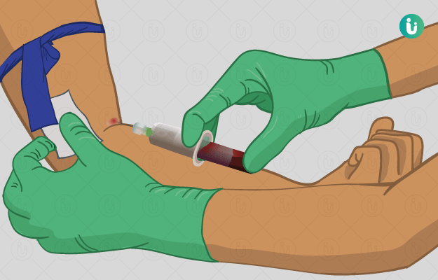
How is a Fatigue Panel test performed?
A blood sample is used to perform the CBC, ESR, blood sugar (random), creatinine, calcium, magnesium, sodium, potassium, chloride, SGPT, TSH and folic acid tests. A technician will draw the required amount of blood from a vein in your arm - multiple samples may be collected for each test.
After the test, you may have a bruise at the needle insertion site that may be painful but will fade over time. You may feel dizzy and faint during or after the test.
The urine R/M test is performed on a urine sample. The urine sample may be collected by either the “clean-catch” method or the 24-hour collection method. The lab will provide you with a special container to collect the sample.
The procedure for the “clean-catch” method is as follows:
- Clean your hands with soap and water.
- Clean your genital area properly.
- Start urinating a little into the toilet bowl and then, stop the flow of urine.
- Now urinate into the container provided until it is about half full.
- Finish urinating into the toilet.
- Close the container and return it to the laboratory for testing.
A 24-hour urine collection is best started early in the morning. The procedure for the 24-hour urine sample collection is as follows:
- On the first day, urinate in the toilet in the morning - the first urine of the day is not collected on day 1.
- After that, collect all the urine in a container for the next 24 hours.
- On the second day, collect your first urine of the morning in the container.
- Seal the sample and return it to the lab.
- Make sure to store the sample in the refrigerator during the collection period.
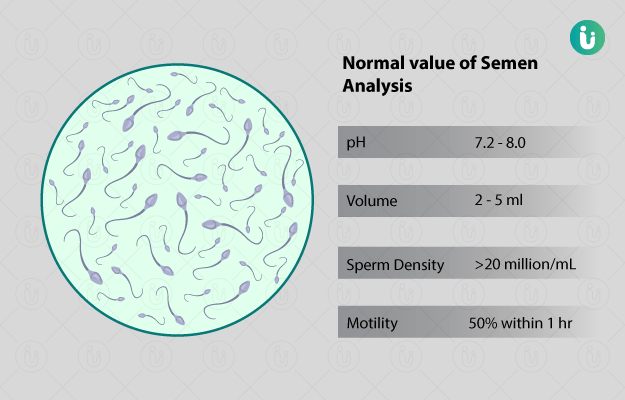
What do Fatigue Panel test results mean?
Normal results:
The normal results of the different tests that are part of the fatigue panel are:
CBC:
- WBC (men and non-pregnant women): 5,000-10,000 WBCs per cubic millimetre (mm3)
- WBC differential:
- Lymphocytes: 25%-40%
- Neutrophils: 50%-62%
- Band neutrophils: 3%-6%
- Monocytes: 3%-7%
- Eosinophils: 0%-3%
- Basophils: 0%-1%
- RBC:
- Men: 4.5-5.5 million RBCs per microlitre (mcL)
- Women: 4.0-5.0 million RBCs/mcL
- Children: 3.8-6.0 million RBCs/mcL
- Newborn: 4.1-6.1 million RBCs/mcL
- Hgb:
- Men: 14-17.4 grams per decilitre (g/dL)
- Women: 12-16 g/dL
- Children: 9.5-20.5 g/dL
- Newborn: 14.5-24.5 g/dL
- HCT:
- Men: 42%-52%
- Women: 36%-48%
- Children: 29%-59%
- Newborn: 44%-64%
- MCH (adults): 28-34 picograms (pg) per cell
- MCHC (adults): 32-36 g/dL
- MCV (adults): 84-96 femtolitres (fL)
- RDW: 11.5%-14.5%
- Platelet:
- Adults: 140,000-400,000 platelets/mm3
- Children: 150,000–450,000 platelets/mm3
- MPV:
- Adults: 7.4-10.4 fL
- Children: 7.4-10.4 fL
- ESR:
- Men (older than 50 years): 0-20 millimetres per hour (mm/hr)
- Men: 0-15 mm/hr
- Women (older than 50 years): 0-30 mm/hr
- Women: 0-20 mm/hr
- Children: 0-10 mm/hr
- Newborn: 0-2 mm/hr
- Random blood sugar:
- Before meal or when waking up: 80-120 milligrams per decilitre (mg/dL)
- Bedtime: 100-140 mg/dL
- Creatinine:
- Men: 0.9-1.3 mg/dL
- Women: 0.6-1.1 mg/dL
- Children (3-18 years): 0.5-1.0 mg/dL
- Children (<3 years): 0.3-0.7 mg/dL
- Calcium: 8.5-10.2 mg/dL
- Magnesium: 1.7-2.2 mg/dL
- Sodium: 135-145 milliequivalents per litre (mEq/L)
- Potassium: 3.7-5.2 mEq/L
- Chloride: 96-106 mEq/L
- SGPT: less than 40 international units per litre (IU/L)
- TSH: 0.5-5 microunits per millilitre (µU/mL)
- Folic acid: 2.7-17.0 nanograms per millilitre (ng/mL)
- Urine R/M test:
- Colour: almost colourless to dark yellow
- Glucose: absent
- Ketones: absent
- Protein: absent
- Bilirubin: absent
- Hgb: absent
- Nitrites: absent
- RBCs: absent
- WBCs: absent
Abnormal results:
The conditions indicated by higher than normal values of different tests are as follows:
-
RBC:
- Polycythaemia vera (a bone marrow disorder)
- Diarrhoea or vomiting
- Dehydration
- Excessive sweating
- Alcohol use disorders
- Cancer
- Liver diseases
- WBC:
- Infection
- Inflammation
- Removal of the spleen
- Rheumatoid arthritis
- Tuberculosis
- Systemic lupus erythematosus (a long-term disease affecting multiple organs)
- Leukaemia (blood cancer)
- Cancer
- Platelets:
- Bleeding
- Iron deficiency
- Cancer
- Differential WBC count:
- Neutrophils:
- Acute suppurative infection (a type of infection with pus formation)
- Trauma (injury to the body)
- Inflammatory disorders (e.g., rheumatic fever, thyroiditis and rheumatoid arthritis)
- Cushing syndrome (a disorder in which there is excessive cortisol production)[44]
- Myelocytic leukaemia (a type of leukaemia)
- Metabolic disorders (conditions in which the body’s metabolism fails, e.g., ketoacidosis, gout and eclampsia)
- Lymphocytes:
- Chronic bacterial infection
- Infectious hepatitis (inflammation of the liver due to an infection)
- Viral infection (e.g., mumps, rubella)
- Multiple myeloma (a cancer of the plasma cells)
- Infectious mononucleosis (an infection caused due to Epstein-Barr virus)
- Lymphocytic leukaemia (a type of leukaemia)
- Radiation
- Monocytes:
- Parasitic infections (e.g., malaria)
- Viral infections (e.g., infectious mononucleosis)
- Chronic inflammatory disorders
- Tuberculosis
- Chronic ulcerative colitis (an immune system disorder that causes inflammation of the lining of the colon)
- Eosinophils:
- Allergic reactions
- Parasitic infections
- Eczema (a group of conditions causing red, itchy and inflamed skin)
- Autoimmune diseases
- Leukaemia
- Basophils:
- Uraemia (a disorder in which waste that should be eliminated through urine is retained in the body)
- Leukaemia
- Myeloproliferative disease (diseases of the bone marrow and blood, e.g., myelofibrosis and polycythaemia rubra vera)
- Neutrophils:
- HCT:
- Burns
- Congenital (present from birth) heart disease
- Severe dehydration
- Polycythaemia vera
- Erythrocytosis (increased production of RBCs)
- Eclampsia (the onset of seizures in pregnant women with high blood pressure)
- Chronic obstructive pulmonary disease
- Hgb:
- Severe burns
- Congenital heart disease
- Haemoconcentration of the blood (increase in RBC and plasma protein due to decrease in the fluid volume in blood)
- Polycythaemia vera
- Chronic obstructive pulmonary disease (obstructive lung disease)
- Congestive heart failure (inability of the heart to pump blood effectively)
- Dehydration
- MCV:
- Liver disease
- Pernicious anaemia (a type of anaemia)
- Alcoholism
- Folic acid deficiency
- MCH:
- Macrocytic anaemia (a type of anaemia)
- MCHC:
- Intravascular haemolysis (destruction of RBCs in circulation in blood)
- Spherocytosis (RBC disorder)
- Cold agglutinins (a rare type of anaemia)
- RDW:
- Iron-deficiency anaemia (anaemia due to iron deficiency)
- B12 or folate-deficiency anaemia (anaemia due to vitamin B12 deficiency)
- Sickle cell disease (a group of disorders that affects the Hgb in the RBCs)
- Haemolytic anaemias
- Posthemorrhagic anaemias (anaemia due to blood loss)
- MPV:
- B12 or folate deficiency
- Valvular heart disease (heart disease affecting the valves in the heart)
- Immune thrombocytopenia (a blood disorder characterised by a deficiency of platelets)
- Massive haemorrhage (loss of large volumes of blood)
- Myelogenous leukaemia (a type of blood cancer)
- ESR:
- Severe stress
- Pregnancy
- Chronic kidney disease
- Infections (pneumonia, pelvic inflammatory disease or appendicitis)
- Polymyalgia rheumatica (inflammation of the joints)
- Graves’ disease (inflammation of the thyroid gland)
- Infection in the kidney, joint, skin or heart valve
- Cancer such as lymphoma or multiple myeloma
- Random blood sugar:
- Type 2 diabetes
- Cushing’s syndrome
- Stroke (loss of blood flow to a part of the brain leading to cell death)
- Acromegaly (excess production of growth hormone)
- Creatinine:
- Dehydration
- Shock (deficient oxygen supply to the cells and tissues caused by failure of blood circulation system)
- Overactive thyroid gland
- Muscle disease
- Blockage in the urinary system
- Congestive heart failure
- Kidney disease
- Calcium:
- Hyperthyroidism
- Hyperparathyroidism (oversecretion of parathyroid hormone)
- Multiple myeloma
- Infections such as TB
- Sarcoidosis (a condition commonly affecting the lungs and skin)
- Paget's disease (chronic skeletal disease)
- Tumours producing a parathyroid hormone-like substance
- Metastatic bone tumour (a cancer that has spread to the bone)
- Magnesium:
- Dehydration
- Adrenal insufficiency (a condition in which the adrenal glands do not produce enough hormones)
- Milk alkali syndrome (high calcium levels in the body which may lead to kidney function loss)
- Diabetic ketoacidosis (the build-up of a harmful substance called ketones in your body in people that have diabetes)
- Acute or chronic kidney failure
- Sodium:
- Excessive use of sodium bicarbonate or salt
- Diarrhoea
- Excessive sweating
- Diabetes insipidus (a type of diabetes in which kidneys are not able to store water)
- Cushing’s syndrome or hyperaldosteronism
- Potassium:
- Blood transfusion
- Excessive potassium in the diet
- Crushed tissue injury (injury caused due to the body part being crushed by a heavy object)
- Metabolic or respiratory acidosis (overproduction of acid or loss of bicarbonate from the blood-metabolic acidosis or the build-up of carbon dioxide in the blood)
- RBC destruction
- Hypoaldosteronism (a condition in which there is a deficiency of aldosterone)
- Hyperkalaemic periodic paralysis (a condition of extreme muscle weakness or paralysis that may begin from infancy)
- Addison’s disease (a rare disorder of the adrenal glands)
- Kidney failure
- Chloride:
- Renal tubular acidosis (excessive acid build-up due to malfunctioning of the kidneys)
- Bromide poisoning
- Metabolic acidosis
- Respiratory alkalosis
- SGPT: Values more than 1,000 IU/L indicate the following:
- Lack of blood flow to the liver
- Acute viral hepatitis (inflammation of liver due to virus)
- Injuries from drugs or toxins
- TSH:
- Hypothyroidism
-
Folic acid:
- Pernicious anaemia (a type of anaemia)
The conditions indicated by lower than normal values of different tests are as follows:
-
RBC:
- Anaemia (due to heavy menstrual bleeding, colon cancer, stomach ulcers, Addison’s disease, sickle-cell disease or lead poisoning)
- Pernicious anaemia (due to the lack of folic acid or vitamin B12)
- WBC:
- Aplastic anaemia (a type of anaemia)
- Viral infections
- Malaria
- Alcoholism
- AIDS
- Systemic lupus erythematosus
- Cushing’s syndrome
- Large spleen
- Platelets:
- Pregnancy
- Large spleen
- Immune thrombocytopenic purpura (decreased platelets in the blood)
- Differential WBC count:
-
Neutrophils:
- Lymphocytes:
- Systemic lupus erythematosus
- Sepsis (a life-threatening response to an infection)
- Leukaemia
- Immunodeficiency diseases (diseases that weaken the immune system’s ability to protect the body)
- Later stages of HIV infection
- Radiation therapy
- Monocytes:
- Hairy-cell leukaemia (a type of blood cancer)
- Aplastic anaemia
- Eosinophils:
- Increased adrenosteroid production
-
Basophils:
- Stress reaction
- Hyperthyroidism
- Acute allergic reactions
-
- HCT:
- Normal pregnancy
- Anaemia
- Hyperthyroidism
- Haemolytic reaction (rupture of RBCs and the leakage of their contents)
- Rheumatoid arthritis
- Haemorrhage (loss of blood from a blood vessel)
- Kidney disease
- Lymphoma (a type of cancer)
- Hodgkin disease (a type of lymphoma)
- Cirrhosis (liver damage leading to scarring)
- Bone marrow failure
- Multiple myeloma
- Hemoglobinopathy
- Prosthetic valves
- Leukaemia
- Hgb:
- Nutritional deficiency
- Anaemia
- Haemorrhage
- Systemic lupus erythematosus
- Chronic haemorrhage (haemorrhage that persists for a long time)
- Splenomegaly (enlargement of the spleen)
- Sarcoidosis
- Haemoglobinopathies (blood disorders like thalassemia and sickle cell disease)
- Kidney disease
- Lymphoma
- Cancer
- Haemolysis (rupture of RBCs and leakage of its contents)
- MCV:
- Iron-deficiency anaemia
- Anaemia due to chronic illness
- Thalassemia (blood disorder)
- MCH:
- Hypochromic anaemia (a type of anaemia)
- Microcytic anaemia (a type of anaemia)
- MCHC:
- Thalassemia
- Iron-deficiency anaemia
- MPV:
- Chemotherapy-induced myelosuppression (decrease in the bone marrow activity of producing WBCs, RBCs and platelets due to cancer therapy)
- Aplastic anaemia
- Wiskott–Aldrich syndrome (a condition associated with lowered ability to form blood clots)
- ESR:
- Sickle-cell disease
- High blood sugar levels
- Polycythaemia
- Liver disease
- Random blood sugar:
- Malnutrition
- Hypothyroidism
- Anorexia (eating disorder)
- Addison’s disease
- Cirrhosis
- Creatinine:
- Muscle loss
- Protein deficiency in the diet
- Severe liver disease
- Calcium:
- Vitamin D deficiency
- Magnesium deficiency
- Osteomalacia (weakening of bones due to lack of vitamin D)
- Hypoparathyroidism (a rare condition in which the parathyroid glands produce very little parathyroid hormone)
- Pancreatitis
- Liver disease
- Kidney failure
- Magnesium:
- Alcohol use disorder
- Chronic diarrhoea
- Hypercalcaemia (high blood calcium level)
- Hyperaldosteronism (excessive production of the hormone, aldosterone, by the adrenal gland)
- Uncontrolled diabetes
- Ulcerative colitis (inflammation of the lining of the large intestine and rectum)
- Kidney disease
- Pancreatitis
- Pre-eclampsia (high blood pressure and the presence of protein in the urine in a pregnant woman)
- Sodium:
- Ketonuria (a condition in which ketone bodies, which are produced due to the burning of fat are found in urine)
- Addison’s disease
- Excessive vasopressin, a type of hormone
- Liver cirrhosis
- Potassium:
- Potassium deficiency in the diet
- Chronic diarrhoea
- Excessive vomiting
- Renal artery stenosis (narrowing of one or both arteries of the kidney)
- Hyperaldosteronism
- Cushing’s syndrome
- Renal tubular acidosis
- Hypokalaemic periodic paralysis (reduced potassium levels in the blood along with muscle weakness)
- Chloride:
- Burns
- Excessive vomiting
- Dehydration
- Excessive sweating
- Bartter syndrome (a group of kidney disorders that cause sodium, potassium and chloride imbalance)
- Syndrome of inappropriate diuretic hormone secretion
- Gastric suction
- Congestive heart failure
- Addison’s disease
- Hyperaldosteronism
- Metabolic alkalosis
- Respiratory acidosis
- TSH:
- Excessive iodine in the body
- Graves’ disease
- Toxic nodular goitre (a condition of excess thyroid hormone production by small masses present in an enlarged thyroid gland)
- Folic acid:
- Malnutrition
- Poor diet
- Malabsorption syndrome (e.g. coeliac sprue)
The folic acid test results may also help in diagnosing conditions such as megaloblastic anaemia and anaemia due to folate deficiency.
Abnormal urine R/M test results indicate the following:
- Kidney stones
- Urinary tract infection
- Bladder or kidney cancer
- Poorly controlled diabetes
Disclaimer: All results must be clinically correlated with the patient’s complaints to make a complete and accurate diagnosis. The above information is provided from a purely educational point of view and is in no way a substitute for medical advice by a qualified doctor.

References
- Better health channel. Department of Health and Human Services [internet]. State government of Victoria; Fatigue
- Michigan Medicine: University of Michigan [internet]. US; Health Library
- University of Rochester Medical Center [Internet]. Rochester (NY): University of Rochester Medical Center; Adult and Children's Health Encyclopedia
- Saha BK, et al. Study of association of primary vesicoureteric reflux in children suffering from urinary tract infection. Chattagram Maa-O-Shishu Hospital Medical College Journal. 2014 Jan;13(1):20-25.
- UF Health [Internet]. University of Florida Health. Florida. US; Tests
- Nemours Children’s Health System [Internet]. Jacksonville (FL): The Nemours Foundation; c2017; For parents
- UCSF health: University of California [internet]. US; Medical Tests
- Benioff Children's Hospital [internet]. University of California. San Francisco. US; Medical Tests
- National Health Service [internet]. UK; Blood Tests
- Goldman L, Schafer AI, eds. Goldman-Cecil Medicine. 25th ed. Philadelphia, PA: Elsevier Saunders; 2016
- Germann CA, Holmes JA. Selected urologic disorders. In: Walls RM, Hockberger RS, Gausche-Hill M. Rosen's Emergency Medicine: Concepts and Clinical Practice. 9th ed. Philadelphia, PA: Elsevier; 2018:chap 89.
- Chau K, Hutton H, Levin A. Laboratory assessment of kidney disease: glomerular filtration rate, urinalysis, and proteinuria. In: Skorecki K, Chertow GM, Marsden PA, Taal MW, Yu ASL, eds. Brenner and Rector's The Kidney. 10th ed. Philadelphia, PA: Elsevier; 2016:chap 26.
- American College of Rheumatology [internet]. Rheumatology Research Foundation. Atlanta. Georgia. U.S.A.; Lupus
- Pagana KD, et al. Mosby’s diagnostic and laboratory test reference. 14th ed. Missouri: Elsevier;2019. White blood cell count and differential count; p.977-978.
- National Institute of Diabetes and Digestive and Kidney Diseases [internet]: US Department of Health and Human Services; Cushing's Syndrome
- American Cancer Society [internet]. Atlanta (GA), USA; What Is Multiple Myeloma?
- Merck Manual Professional Version [Internet]. Kenilworth (NJ): Merck & Co. Inc.; c2019. Infectious Mononucleosis
- Virginia Mason Medical Center [Internet]. Seattle. US; Colitis and Chronic Ulcerative Colitis
- National Eczema Association [Internet]. California. US; What is Eczema?
- MD Anderson Cancer Center: The University of Texas [Internet]. US; Myeloproliferative Disorder
- Meyer TW and Hostetter TH. Approaches to uremia. JASN. 2014 Oct; 25(10):2151-2158.
- Merck Manual Consumer Version [Internet]. Kenilworth (NJ): Merck & Co. Inc.; c2018. Erythrocytosis
- Merck Manual Professional Version [Internet]. Kenilworth (NJ): Merck & Co. Inc.; c2019. Preeclampsia and Eclampsia
- American Heart Association [internet]. Dallas. Texas. U.S.A.; Congestive Heart Failure and Congenital Defects
- Dhaliwal G, et al. Hemolytic anemia. Am Fam Physician. 2004 Jun 1;69(11):2599-2607. PMID: 15202694.
- Beth Israel Lahey Health: Winchester Hospital [Internet]. Winchester. Maryland. US; Health Library.
- Genetic and Rare Diseases Information Center. National Center for Advancing Translational Sciences. National Institute of Health. U.S. Department of Health and Human Services; Cold agglutinin disease
- Genetics Home Reference [internet]. National Institute of Health: US National Library of Medicine. US Department of Health and Human Services; Sickle cell disease
- Kirkman HN and Riley HD. Posthemorrhagic anemia and shock in the newborn- A Review. Pediatrics July 1959, 24(1);97-105. PMID: 13667339.
- Joint United Kingdom (UK) Blood Transfusion and Tissue Transplantation Services Professional Advisory Committee [Internet]. UK; 7.3: Transfusion management of major haemorrhage.
- Genetics Home Reference [internet]. National Institute of Health: US National Library of Medicine. US Department of Health and Human Services; Immune thrombocytopenia
- National Health Service [internet]. UK; Sarcoidosis
- Haseer Koya H, Paul M. Shock. [Updated 2020 Jan 28]. In: StatPearls [Internet]. Treasure Island (FL): StatPearls Publishing; 2020 Jan
- National Cancer Institute [Internet]. Bethesda (MD): U.S. Department of Health and Human Services; NCI Dictionary of Cancer Terms
- American Academy of Orthopaedic Surgeons [Internet]. Illinois. US; Paget's Disease of Bone
- Centers for Disease Control and Prevention [internet]. Atlanta (GA): US Department of Health and Human Services; What is sepsis?
- Harewood J, Master SR. Hemolytic Transfusion Reaction. [Updated 2019 Jun 4]. In: StatPearls [Internet]. Treasure Island (FL): StatPearls Publishing; 2020 Jan
- UCLA health [Internet]. University of California. Oakland. California. US; Toxic Nodular Goiter
- Comstock JP, Garber AJ. Ketonuria. In: Walker HK, Hall WD, Hurst JW, editors. Clinical Methods: The History, Physical, and Laboratory Examinations. 3rd edition. Boston: Butterworths; 1990. Chapter 140.